3OAX

Crystal structure of bovine rhodopsin with beta-ionone


Experimental Data Snapshot

  • Method: X-RAY DIFFRACTION
  • Resolution: 2.60 Å
  • R-Value Free: 0.262 
  • R-Value Work: 0.224 
  • R-Value Observed: 0.224 

wwPDB Validation   3D Report Full Report


Ligand Structure Quality Assessment 


This is version 2.1 of the entry. See complete history


Literature

Binding of more than one retinoid to visual opsins

Makino, C.L.Riley, C.K.Looney, J.Crouch, R.K.Okada, T.

(2010) Biophys J 99: 2366-2373

  • DOI: https://doi.org/10.1016/j.bpj.2010.08.003
  • Primary Citation of Related Structures:  
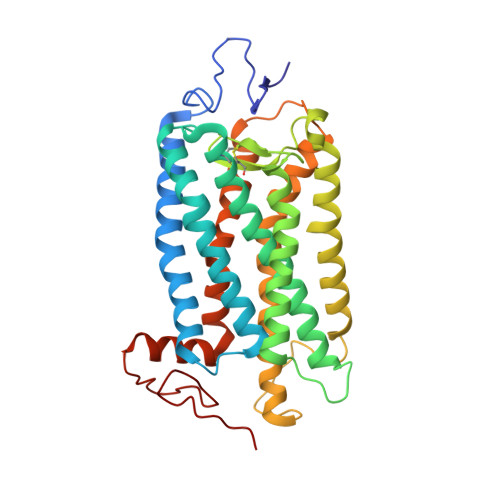
    3OAX

  • PubMed Abstract: 

    Visual opsins bind 11-cis retinal at an orthosteric site to form rhodopsins but increasing evidence suggests that at least some are capable of binding an additional retinoid(s) at a separate, allosteric site(s). Microspectrophotometric measurements on isolated, dark-adapted, salamander photoreceptors indicated that the truncated retinal analog, β-ionone, partitioned into the membranes of green-sensitive rods; however, in blue-sensitive rod outer segments, there was an enhanced uptake of four or more β-ionones per rhodopsin. X-ray crystallography revealed binding of one β-ionone to bovine green-sensitive rod rhodopsin. Cocrystallization only succeeded with extremely high concentrations of β-ionone and binding did not alter the structure of rhodopsin from the inactive state. Salamander green-sensitive rod rhodopsin is also expected to bind β-ionone at sufficiently high concentrations because the binding site is present on its surface. Therefore, both blue- and green-sensitive rod rhodopsins have at least one allosteric binding site for retinoid, but β-ionone binds to the latter type of rhodopsin with low affinity and low efficacy.


  • Organizational Affiliation

    Department of Ophthalmology, Massachusetts Eye and Ear Infirmary, Boston, MA, USA. cmakino@meei.harvard.edu


Macromolecules
Find similar proteins by:  (by identity cutoff)  |  3D Structure
Entity ID: 1
MoleculeChains Sequence LengthOrganismDetailsImage
Rhodopsin
A, B
349Bos taurusMutation(s): 1 
Membrane Entity: Yes 
UniProt
Find proteins for P02699 (Bos taurus)
Explore P02699 
Go to UniProtKB:  P02699
Entity Groups  
Sequence Clusters30% Identity50% Identity70% Identity90% Identity95% Identity100% Identity
UniProt GroupP02699
Sequence Annotations
Expand
  • Reference Sequence
Oligosaccharides

Help

Entity ID: 2
MoleculeChains Length2D Diagram Glycosylation3D Interactions
beta-D-mannopyranose-(1-4)-2-acetamido-2-deoxy-beta-D-glucopyranose-(1-4)-2-acetamido-2-deoxy-beta-D-glucopyranose
C
3N-Glycosylation
Glycosylation Resources
GlyTouCan:  G15407YE
GlyCosmos:  G15407YE
GlyGen:  G15407YE
Entity ID: 3
MoleculeChains Length2D Diagram Glycosylation3D Interactions
2-acetamido-2-deoxy-beta-D-glucopyranose-(1-4)-2-acetamido-2-deoxy-beta-D-glucopyranose
D, F
2N-Glycosylation
Glycosylation Resources
GlyTouCan:  G42666HT
GlyCosmos:  G42666HT
GlyGen:  G42666HT
Entity ID: 4
MoleculeChains Length2D Diagram Glycosylation3D Interactions
alpha-D-mannopyranose-(1-3)-beta-D-mannopyranose-(1-4)-2-acetamido-2-deoxy-beta-D-glucopyranose-(1-4)-2-acetamido-2-deoxy-beta-D-glucopyranose
E
4N-Glycosylation
Glycosylation Resources
GlyTouCan:  G81315DD
GlyCosmos:  G81315DD
GlyGen:  G81315DD
Small Molecules
Ligands 8 Unique
IDChains Name / Formula / InChI Key2D Diagram3D Interactions
HTG
Query on HTG

Download Ideal Coordinates CCD File 
FA [auth B],
GA [auth B],
R [auth A],
S [auth A]
heptyl 1-thio-beta-D-glucopyranoside
C13 H26 O5 S
HPEGNLMTTNTJSP-LBELIVKGSA-N
RET
Query on RET

Download Ideal Coordinates CCD File 
G [auth A],
U [auth B]
RETINAL
C20 H28 O
NCYCYZXNIZJOKI-OVSJKPMPSA-N
PLM
Query on PLM

Download Ideal Coordinates CCD File 
H [auth A],
I [auth A],
V [auth B],
W [auth B]
PALMITIC ACID
C16 H32 O2
IPCSVZSSVZVIGE-UHFFFAOYSA-N
4E6
Query on 4E6

Download Ideal Coordinates CCD File 
EA [auth B],
Q [auth A]
(4E,6E)-hexadeca-1,4,6-triene
C16 H28
OPBISVRDVDRVHM-PTSVWTKZSA-N
HG
Query on HG

Download Ideal Coordinates CCD File 
J [auth A]
K [auth A]
L [auth A]
X [auth B]
Y [auth B]
J [auth A],
K [auth A],
L [auth A],
X [auth B],
Y [auth B],
Z [auth B]
MERCURY (II) ION
Hg
BQPIGGFYSBELGY-UHFFFAOYSA-N
ID3
Query on ID3

Download Ideal Coordinates CCD File 
HA [auth B],
T [auth A]
(3E)-4-(2,6,6-trimethylcyclohex-1-en-1-yl)but-3-en-2-one
C13 H20 O
PSQYTAPXSHCGMF-BQYQJAHWSA-N
HTO
Query on HTO

Download Ideal Coordinates CCD File 
DA [auth B]HEPTANE-1,2,3-TRIOL
C7 H16 O3
HXYCHJFUBNTKQR-RNFRBKRXSA-N
ZN
Query on ZN

Download Ideal Coordinates CCD File 
AA [auth B]
BA [auth B]
CA [auth B]
M [auth A]
N [auth A]
AA [auth B],
BA [auth B],
CA [auth B],
M [auth A],
N [auth A],
O [auth A],
P [auth A]
ZINC ION
Zn
PTFCDOFLOPIGGS-UHFFFAOYSA-N
Experimental Data & Validation

Experimental Data

  • Method: X-RAY DIFFRACTION
  • Resolution: 2.60 Å
  • R-Value Free: 0.262 
  • R-Value Work: 0.224 
  • R-Value Observed: 0.224 
  • Space Group: P 41
Unit Cell:
Length ( Å )Angle ( ˚ )
a = 96.99α = 90
b = 96.99β = 90
c = 149.8γ = 90
Software Package:
Software NamePurpose
ADSCdata collection
AMoREphasing
CNSrefinement
HKL-2000data reduction
SCALEPACKdata scaling

Structure Validation

View Full Validation Report


Ligand Structure Quality Assessment 


Entry History 

Deposition Data

Revision History  (Full details and data files)

  • Version 1.0: 2011-01-19
    Type: Initial release
  • Version 1.1: 2011-07-13
    Changes: Version format compliance
  • Version 2.0: 2020-07-29
    Type: Remediation
    Reason: Carbohydrate remediation
    Changes: Atomic model, Data collection, Database references, Derived calculations, Structure summary
  • Version 2.1: 2023-11-01
    Changes: Data collection, Database references, Refinement description, Structure summary