


Crystal structure of an ADP-ribose pyrophosphatase (SSU98_1448) from STREPTOCOCCUS SUIS 89-1591 at 2.27 A resolution
Joint Center for Structural Genomics (JCSG)To be published.
Experimental Data Snapshot
wwPDB Validation 3D Report Full Report

Entity ID: 1 | |||||
|---|---|---|---|---|---|
| Molecule | Chains | Sequence Length | Organism | Details | Image |
| ADP-ribose pyrophosphatase | 206 | Streptococcus suis 89/1591 | Mutation(s): 0 Gene Names: SsuiDRAFT_3457 | 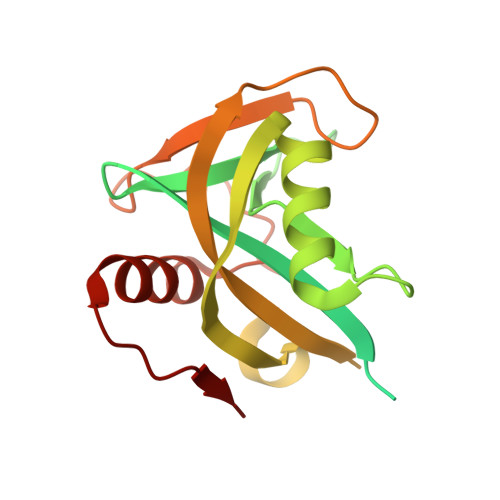 | |
Entity Groups | |||||
| Sequence Clusters | 30% Identity50% Identity70% Identity90% Identity95% Identity100% Identity | ||||
Sequence AnnotationsExpand | |||||
| |||||
| Ligands 2 Unique | |||||
|---|---|---|---|---|---|
| ID | Chains | Name / Formula / InChI Key | 2D Diagram | 3D Interactions | |
| EDO Query on EDO | B [auth A], C [auth A], D [auth A], E [auth A], F [auth A] | 1,2-ETHANEDIOL C2 H6 O2 LYCAIKOWRPUZTN-UHFFFAOYSA-N |  | ||
| ACT Query on ACT | G [auth A] | ACETATE ION C2 H3 O2 QTBSBXVTEAMEQO-UHFFFAOYSA-M |  | ||
| Modified Residues 1 Unique | |||||
|---|---|---|---|---|---|
| ID | Chains | Type | Formula | 2D Diagram | Parent |
| MSE Query on MSE | A | L-PEPTIDE LINKING | C5 H11 N O2 Se |  | MET |
| Length ( Å ) | Angle ( ˚ ) |
|---|---|
| a = 64.78 | α = 90 |
| b = 64.78 | β = 90 |
| c = 108.29 | γ = 90 |
| Software Name | Purpose |
|---|---|
| BUSTER-TNT | refinement |
| XSCALE | data processing |
| PDB_EXTRACT | data extraction |
| XDS | data reduction |
| XSCALE | data scaling |
| Rosetta | phasing |
| PHASER | phasing |
| BUSTER | refinement |
RCSB PDB (citation) is hosted by
RCSB PDB is a member of the

