3O8O

Structure of phosphofructokinase from Saccharomyces cerevisiae


Experimental Data Snapshot

  • Method: X-RAY DIFFRACTION
  • Resolution: 2.90 Å
  • R-Value Free: 0.309 
  • R-Value Work: 0.259 
  • R-Value Observed: 0.259 

wwPDB Validation   3D Report Full Report


Ligand Structure Quality Assessment 


This is version 1.4 of the entry. See complete history


Literature

The Crystal Structures of Eukaryotic Phosphofructokinases from Baker's Yeast and Rabbit Skeletal Muscle.

Banaszak, K.Mechin, I.Obmolova, G.Oldham, M.Chang, S.H.Ruiz, T.Radermacher, M.Kopperschlager, G.Rypniewski, W.

(2011) J Mol Biol 407: 284-297

  • DOI: https://doi.org/10.1016/j.jmb.2011.01.019
  • Primary Citation of Related Structures:  
    3O8L, 3O8N, 3O8O

  • PubMed Abstract: 

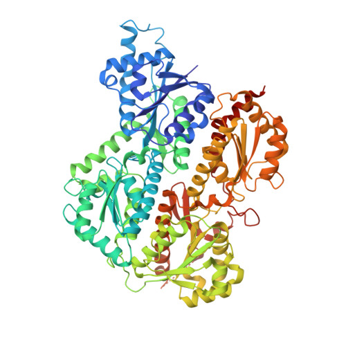
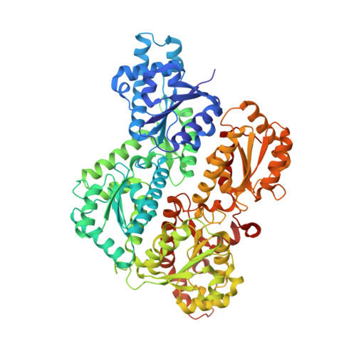
    Phosphofructokinase 1 (PFK) is a multisubunit allosteric enzyme that catalyzes the principal regulatory step in glycolysis-the phosphorylation of fructose 6-phosphate to fructose 1,6-bisphosphate by ATP. The activity of eukaryotic PFK is modulated by a number of effectors in response to the cell's needs for energy and building blocks for biosynthesis. The crystal structures of eukaryotic PFKs-from Saccharomyces cerevisiae and rabbit skeletal muscle-demonstrate how successive gene duplications and fusion are reflected in the protein structure and how they allowed the evolution of new functionalities. The basic framework inherited from prokaryotes is conserved, and additional levels of structural and functional complexity have evolved around it. Analysis of protein-ligand complexes has shown how PFK is activated by fructose 2,6-bisphosphate (a powerful PFK effector found only in eukaryotes) and reveals a novel nucleotide binding site. Crystallographic results have been used as the basis for structure-based effector design.


  • Organizational Affiliation

    Institute of Bioorganic Chemistry, Polish Academy of Sciences, Poznan, Poland.


Macromolecules
Find similar proteins by:  (by identity cutoff)  |  3D Structure
Entity ID: 1
MoleculeChains Sequence LengthOrganismDetailsImage
6-phosphofructokinase subunit alpha
A, C, E, G
787Saccharomyces cerevisiaeMutation(s): 0 
Gene Names: PFK1YGR240CG8599
EC: 2.7.1.11
UniProt
Find proteins for P16861 (Saccharomyces cerevisiae (strain ATCC 204508 / S288c))
Explore P16861 
Go to UniProtKB:  P16861
Entity Groups  
Sequence Clusters30% Identity50% Identity70% Identity90% Identity95% Identity100% Identity
UniProt GroupP16861
Sequence Annotations
Expand
  • Reference Sequence
Find similar proteins by:  (by identity cutoff)  |  3D Structure
Entity ID: 2
MoleculeChains Sequence LengthOrganismDetailsImage
6-phosphofructokinase subunit beta
B, D, F, H
766Saccharomyces cerevisiaeMutation(s): 0 
Gene Names: PFK2YMR205CYM8325.06C
EC: 2.7.1.11
UniProt
Find proteins for P16862 (Saccharomyces cerevisiae (strain ATCC 204508 / S288c))
Explore P16862 
Go to UniProtKB:  P16862
Entity Groups  
Sequence Clusters30% Identity50% Identity70% Identity90% Identity95% Identity100% Identity
UniProt GroupP16862
Sequence Annotations
Expand
  • Reference Sequence
Small Molecules
Ligands 2 Unique
IDChains Name / Formula / InChI Key2D Diagram3D Interactions
FDP
Query on FDP

Download Ideal Coordinates CCD File 
J [auth A]
L [auth B]
N [auth C]
P [auth D]
R [auth E]
J [auth A],
L [auth B],
N [auth C],
P [auth D],
R [auth E],
T [auth F],
V [auth G],
X [auth H]
2,6-di-O-phosphono-beta-D-fructofuranose
C6 H14 O12 P2
YXWOAJXNVLXPMU-ZXXMMSQZSA-N
F6P
Query on F6P

Download Ideal Coordinates CCD File 
I [auth A]
K [auth B]
M [auth C]
O [auth D]
Q [auth E]
I [auth A],
K [auth B],
M [auth C],
O [auth D],
Q [auth E],
S [auth F],
U [auth G],
W [auth H]
6-O-phosphono-beta-D-fructofuranose
C6 H13 O9 P
BGWGXPAPYGQALX-ARQDHWQXSA-N
Experimental Data & Validation

Experimental Data

  • Method: X-RAY DIFFRACTION
  • Resolution: 2.90 Å
  • R-Value Free: 0.309 
  • R-Value Work: 0.259 
  • R-Value Observed: 0.259 
  • Space Group: P 21 21 21
Unit Cell:
Length ( Å )Angle ( ˚ )
a = 180.046α = 90
b = 186.205β = 90
c = 236.507γ = 90
Software Package:
Software NamePurpose
MAR345data collection
AMoREphasing
CNSrefinement
DENZOdata reduction
SCALEPACKdata scaling

Structure Validation

View Full Validation Report


Ligand Structure Quality Assessment 


Entry History 

Deposition Data

Revision History  (Full details and data files)

  • Version 1.0: 2011-02-02
    Type: Initial release
  • Version 1.1: 2011-07-13
    Changes: Version format compliance
  • Version 1.2: 2017-11-08
    Changes: Advisory, Refinement description
  • Version 1.3: 2020-07-29
    Type: Remediation
    Reason: Carbohydrate remediation
    Changes: Data collection, Derived calculations, Structure summary
  • Version 1.4: 2023-09-06
    Changes: Advisory, Data collection, Database references, Refinement description, Structure summary