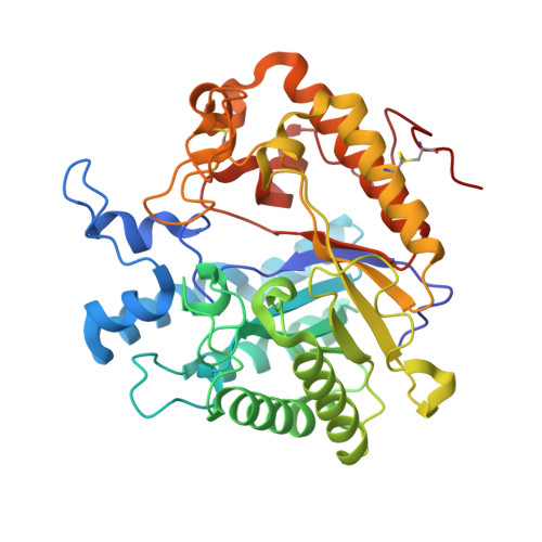
Carbohydrate binding sites in Candida albicans exo-beta-1,3-glucanase and the role of the Phe-Phe 'clamp' at the active site entrance
Patrick, W.M., Nakatani, Y., Cutfield, S.M., Sharpe, M.L., Ramsay, R.J., Cutfield, J.F.(2010) FEBS J 277: 4549-4561
- PubMed: 20875088
- DOI: https://doi.org/10.1111/j.1742-4658.2010.07869.x
- Primary Citation of Related Structures:
2PF0, 3O6A - PubMed Abstract:
Candida albicans exo-β-1,3-glucanase (Exg; EC 3.2.1.58) is implicated in cell wall β-D-glucan remodelling through its glucosyl hydrolase and/or transglucosylase activities. A pair of antiparallel phenylalanyl residues (F144 and F258) flank the entrance to the active site pocket. Various Exg mutants were studied using steady-state kinetics and crystallography aiming to understand the roles played by these residues in positioning the β-1,3-D-glucan substrate. Mutations at the Phe-Phe entranceway demonstrated the requirement for double-sided CH/π interactions at the +1 subsite, and the necessity for phenylalanine rather than tyrosine or tryptophan. The Tyr-Tyr double mutations introduced ordered water molecules into the entranceway. A third Phe residue (F229) nearby was evaluated as a possible +2 subsite. The inactive double mutant E292S/F229A complexed with laminaritriose has provided the first picture of substrate binding to Exg and demonstrated how the Phe-Phe arrangement acts as a clamp at the +1 subsite. The terminal sugar at the -1 site showed displacement from the position of a monosaccharide analogue with interchange of water molecules and sugar hydroxyls. An unexpected additional glucose binding site, well removed from the active site, was revealed. This site may enable Exg to associate with the branched glucan structure of the C. albicans cell wall.
Organizational Affiliation:
Department of Biochemistry, University of Otago, Dunedin, New Zealand.


