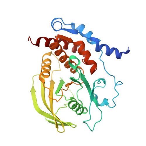
Visualizing active-site dynamics in single crystals of HePTP: opening of the WPD loop involves coordinated movement of the E loop.
Critton, D.A., Tautz, L., Page, R.(2011) J Mol Biol 405: 619-629
- PubMed: 21094165
- DOI: https://doi.org/10.1016/j.jmb.2010.11.020
- Primary Citation of Related Structures:
3O4S, 3O4T, 3O4U - PubMed Abstract:
Phosphotyrosine hydrolysis by protein tyrosine phosphatases (PTPs) involves substrate binding by the PTP loop and closure over the active site by the WPD loop. The E loop, located immediately adjacent to the PTP and WPD loops, is conserved among human PTPs in both sequence and structure, yet the role of this loop in substrate binding and catalysis is comparatively unexplored. Hematopoietic PTP (HePTP) is a member of the kinase interaction motif (KIM) PTP family. Compared to other PTPs, KIM-PTPs have E loops that are unique in both sequence and structure. In order to understand the role of the E loop in the transition between the closed state and the open state of HePTP, we identified a novel crystal form of HePTP that allowed the closed-state-to-open-state transition to be observed within a single crystal form. These structures, which include the first structure of the HePTP open state, show that the WPD loop adopts an 'atypically open' conformation and, importantly, that ligands can be exchanged at the active site, which is critical for HePTP inhibitor development. These structures also show that tetrahedral oxyanions bind at a novel secondary site and function to coordinate the PTP, WPD, and E loops. Finally, using both structural and kinetic data, we reveal a novel role for E-loop residue Lys182 in enhancing HePTP catalytic activity through its interaction with Asp236 of the WPD loop, providing the first evidence for the coordinated dynamics of the WPD and E loops in the catalytic cycle, which, as we show, is relevant to multiple PTP families.
Organizational Affiliation:
Department of Molecular Biology, Cell Biology, and Biochemistry, Brown University, Providence, RI 02912, USA.