3O4P

DFPase at 0.85 Angstrom resolution (H atoms included)


Experimental Data Snapshot

  • Method: X-RAY DIFFRACTION
  • Resolution: 0.85 Å
  • R-Value Free: 0.121 
  • R-Value Work: 0.103 

wwPDB Validation   3D Report Full Report


This is version 1.3 of the entry. See complete history


Literature

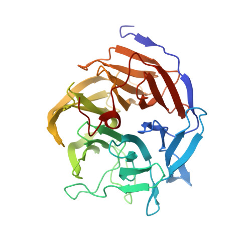
Hydrogen atoms in protein structures: high-resolution X-ray diffraction structure of the DFPase.

Elias, M.Liebschner, D.Koepke, J.Lecomte, C.Guillot, B.Jelsch, C.Chabriere, E.

(2013) BMC Res Notes 6: 308-308

  • DOI: https://doi.org/10.1186/1756-0500-6-308
  • Primary Citation of Related Structures:  
    3O4P

  • PubMed Abstract: 

    Hydrogen atoms represent about half of the total number of atoms in proteins and are often involved in substrate recognition and catalysis. Unfortunately, X-ray protein crystallography at usual resolution fails to access directly their positioning, mainly because light atoms display weak contributions to diffraction. However, sub-Ångstrom diffraction data, careful modeling and a proper refinement strategy can allow the positioning of a significant part of hydrogen atoms.


  • Organizational Affiliation

    Weizmann Institute of Science, Biological Chemistry, Rehovot, Israel.


Macromolecules
Find similar proteins by:  (by identity cutoff)  |  3D Structure
Entity ID: 1
MoleculeChains Sequence LengthOrganismDetailsImage
Diisopropyl-fluorophosphatase314Loligo vulgarisMutation(s): 0 
EC: 3.1.8.2
UniProt
Find proteins for Q7SIG4 (Loligo vulgaris)
Explore Q7SIG4 
Go to UniProtKB:  Q7SIG4
Entity Groups  
Sequence Clusters30% Identity50% Identity70% Identity90% Identity95% Identity100% Identity
UniProt GroupQ7SIG4
Sequence Annotations
Expand
  • Reference Sequence
Small Molecules
Ligands 9 Unique
IDChains Name / Formula / InChI Key2D Diagram3D Interactions
MES
Query on MES

Download Ideal Coordinates CCD File 
D [auth A],
E [auth A]
2-(N-MORPHOLINO)-ETHANESULFONIC ACID
C6 H13 N O4 S
SXGZJKUKBWWHRA-UHFFFAOYSA-N
PGE
Query on PGE

Download Ideal Coordinates CCD File 
N [auth A],
O [auth A]
TRIETHYLENE GLYCOL
C6 H14 O4
ZIBGPFATKBEMQZ-UHFFFAOYSA-N
ME2
Query on ME2

Download Ideal Coordinates CCD File 
V [auth A]1-ETHOXY-2-(2-METHOXYETHOXY)ETHANE
C7 H16 O3
CNJRPYFBORAQAU-UHFFFAOYSA-N
PEG
Query on PEG

Download Ideal Coordinates CCD File 
T [auth A],
U [auth A]
DI(HYDROXYETHYL)ETHER
C4 H10 O3
MTHSVFCYNBDYFN-UHFFFAOYSA-N
GOL
Query on GOL

Download Ideal Coordinates CCD File 
B [auth A],
C [auth A]
GLYCEROL
C3 H8 O3
PEDCQBHIVMGVHV-UHFFFAOYSA-N
DXE
Query on DXE

Download Ideal Coordinates CCD File 
P [auth A],
Q [auth A]
1,2-DIMETHOXYETHANE
C4 H10 O2
XTHFKEDIFFGKHM-UHFFFAOYSA-N
MXE
Query on MXE

Download Ideal Coordinates CCD File 
R [auth A],
S [auth A]
2-METHOXYETHANOL
C3 H8 O2
XNWFRZJHXBZDAG-UHFFFAOYSA-N
EDO
Query on EDO

Download Ideal Coordinates CCD File 
F [auth A]
G [auth A]
H [auth A]
I [auth A]
J [auth A]
F [auth A],
G [auth A],
H [auth A],
I [auth A],
J [auth A],
K [auth A],
L [auth A],
M [auth A]
1,2-ETHANEDIOL
C2 H6 O2
LYCAIKOWRPUZTN-UHFFFAOYSA-N
CA
Query on CA

Download Ideal Coordinates CCD File 
W [auth A],
X [auth A]
CALCIUM ION
Ca
BHPQYMZQTOCNFJ-UHFFFAOYSA-N
Experimental Data & Validation

Experimental Data

  • Method: X-RAY DIFFRACTION
  • Resolution: 0.85 Å
  • R-Value Free: 0.121 
  • R-Value Work: 0.103 
  • Space Group: P 21 21 21
Unit Cell:
Length ( Å )Angle ( ˚ )
a = 43.114α = 90
b = 81.849β = 90
c = 86.467γ = 90
Software Package:
Software NamePurpose
MoProrefinement
MOSFLMdata reduction
SCALAdata scaling

Structure Validation

View Full Validation Report


Entry History 

Deposition Data

Revision History  (Full details and data files)

  • Version 1.0: 2011-08-17
    Type: Initial release
  • Version 1.1: 2013-08-14
    Changes: Database references
  • Version 1.2: 2024-02-21
    Changes: Data collection, Database references, Derived calculations
  • Version 1.3: 2024-04-03
    Changes: Refinement description