3O0Z

Crystal structure of a coiled-coil domain from human ROCK I


Experimental Data Snapshot

  • Method: X-RAY DIFFRACTION
  • Resolution: 2.33 Å
  • R-Value Free: 0.286 
  • R-Value Work: 0.239 
  • R-Value Observed: 0.242 

wwPDB Validation   3D Report Full Report


This is version 1.1 of the entry. See complete history


Literature

Crystal Structure of a Coiled-Coil Domain from Human ROCK I.

Tu, D.Li, Y.Song, H.K.Toms, A.V.Gould, C.J.Ficarro, S.B.Marto, J.A.Goode, B.L.Eck, M.J.

(2011) PLoS One 6: e18080-e18080

  • DOI: https://doi.org/10.1371/journal.pone.0018080
  • Primary Citation of Related Structures:  
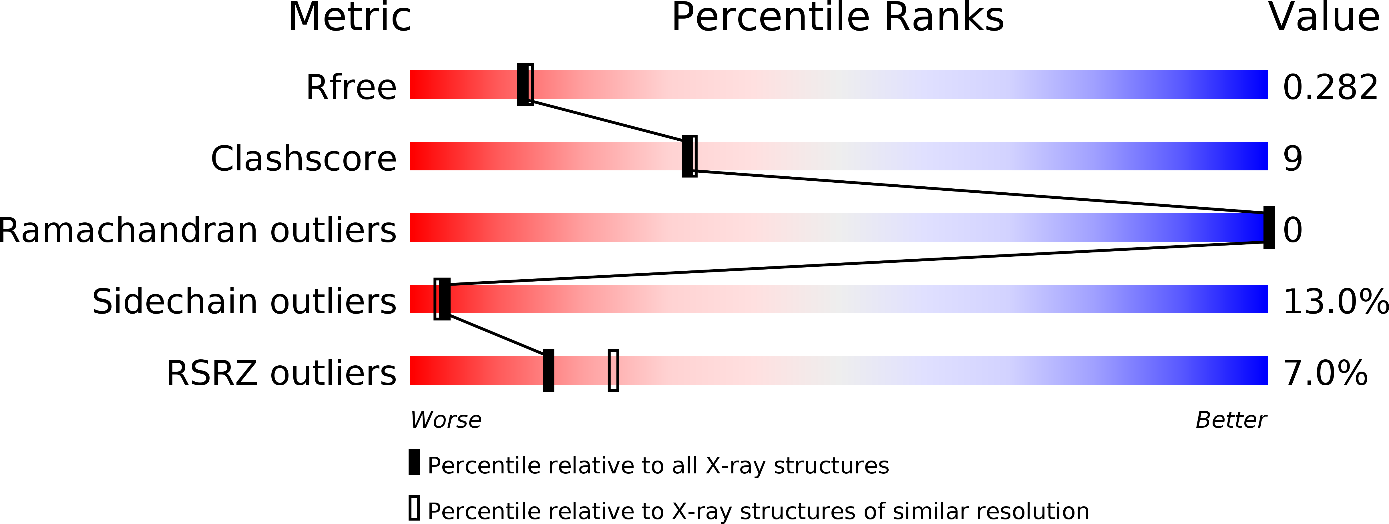
    3O0Z

  • PubMed Abstract: 

    The small GTPase Rho and one of its targets, Rho-associated kinase (ROCK), participate in a variety of actin-based cellular processes including smooth muscle contraction, cell migration, and stress fiber formation. The ROCK protein consists of an N-terminal kinase domain, a central coiled-coil domain containing a Rho binding site, and a C-terminal pleckstrin homology domain. Here we present the crystal structure of a large section of the central coiled-coil domain of human ROCK I (amino acids 535-700). The structure forms a parallel α-helical coiled-coil dimer that is structurally similar to tropomyosin, an actin filament binding protein. There is an unusual discontinuity in the coiled-coil; three charged residues (E613, R617 and D620) are positioned at what is normally the hydrophobic core of coiled-coil packing. We speculate that this conserved irregularity could function as a hinge that allows ROCK to adopt its autoinhibited conformation.


  • Organizational Affiliation

    Department of Biological Chemistry and Molecular Pharmacology, Harvard Medical School, Boston, Massachusetts, United States of America.


Macromolecules
Find similar proteins by:  (by identity cutoff)  |  3D Structure
Entity ID: 1
MoleculeChains Sequence LengthOrganismDetailsImage
Rho-associated protein kinase 1
A, B, C, D
168Homo sapiensMutation(s): 0 
Gene Names: ROCK1
EC: 2.7.11.1
UniProt & NIH Common Fund Data Resources
Find proteins for Q13464 (Homo sapiens)
Explore Q13464 
Go to UniProtKB:  Q13464
PHAROS:  Q13464
GTEx:  ENSG00000067900 
Entity Groups  
Sequence Clusters30% Identity50% Identity70% Identity90% Identity95% Identity100% Identity
UniProt GroupQ13464
Sequence Annotations
Expand
  • Reference Sequence
Small Molecules
Modified Residues  1 Unique
IDChains TypeFormula2D DiagramParent
MSE
Query on MSE
A, B, C, D
L-PEPTIDE LINKINGC5 H11 N O2 SeMET
Experimental Data & Validation

Experimental Data

  • Method: X-RAY DIFFRACTION
  • Resolution: 2.33 Å
  • R-Value Free: 0.286 
  • R-Value Work: 0.239 
  • R-Value Observed: 0.242 
  • Space Group: P 43 21 2
Unit Cell:
Length ( Å )Angle ( ˚ )
a = 84.588α = 90
b = 84.588β = 90
c = 340.129γ = 90
Software Package:
Software NamePurpose
HKL-2000data collection
PHASERphasing
PHENIXrefinement
HKL-2000data reduction
HKL-2000data scaling

Structure Validation

View Full Validation Report


Entry History 

Deposition Data

Revision History  (Full details and data files)

  • Version 1.0: 2011-03-23
    Type: Initial release
  • Version 1.1: 2011-07-13
    Changes: Version format compliance