3O0J

PDE4B In complex with ligand an2898


Experimental Data Snapshot

  • Method: X-RAY DIFFRACTION
  • Resolution: 1.95 Å
  • R-Value Free: 0.236 
  • R-Value Work: 0.184 
  • R-Value Observed: 0.187 

wwPDB Validation   3D Report Full Report


Ligand Structure Quality Assessment 


This is version 1.3 of the entry. See complete history


Literature

Boron-based phosphodiesterase inhibitors show novel binding of boron to PDE4 bimetal center.

Freund, Y.R.Akama, T.Alley, M.R.Antunes, J.Dong, C.Jarnagin, K.Kimura, R.Nieman, J.A.Maples, K.R.Plattner, J.J.Rock, F.Sharma, R.Singh, R.Sanders, V.Zhou, Y.

(2012) FEBS Lett 586: 3410-3414

  • DOI: https://doi.org/10.1016/j.febslet.2012.07.058
  • Primary Citation of Related Structures:  
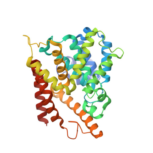
    3O0J

  • PubMed Abstract: 

    We have used boron-based molecules to create novel, competitive, reversible inhibitors of phosphodiesterase 4 (PDE4). The co-crystal structure reveals a binding configuration which is unique compared to classical catechol PDE4 inhibitors, with boron binding to the activated water in the bimetal center. These phenoxybenzoxaboroles can be optimized to generate submicromolar potency enzyme inhibitors, which inhibit TNF-α, IL-2, IFN-γ, IL-5 and IL-10 activities in vitro and show safety and efficacy for topical treatment of human psoriasis. They provide a valuable new route for creating novel potent anti-PDE4 inhibitors.


  • Organizational Affiliation

    Anacor Pharmaceuticals Inc., Palo Alto, CA 94303, USA. yfreund@anacor.com


Macromolecules
Find similar proteins by:  (by identity cutoff)  |  3D Structure
Entity ID: 1
MoleculeChains Sequence LengthOrganismDetailsImage
cAMP-specific 3',5'-cyclic phosphodiesterase 4B323Homo sapiensMutation(s): 0 
EC: 3.1.4.17
UniProt & NIH Common Fund Data Resources
Find proteins for Q07343 (Homo sapiens)
Explore Q07343 
Go to UniProtKB:  Q07343
PHAROS:  Q07343
GTEx:  ENSG00000184588 
Entity Groups  
Sequence Clusters30% Identity50% Identity70% Identity90% Identity95% Identity100% Identity
UniProt GroupQ07343
Sequence Annotations
Expand
  • Reference Sequence
Small Molecules
Ligands 4 Unique
IDChains Name / Formula / InChI Key2D Diagram3D Interactions
3OJ
Query on 3OJ

Download Ideal Coordinates CCD File 
D [auth A]4-[(1-hydroxy-1,3-dihydro-2,1-benzoxaborol-5-yl)oxy]benzene-1,2-dicarbonitrile
C15 H9 B N2 O3
UBMGTTRDNUKZMT-UHFFFAOYSA-N
ZN
Query on ZN

Download Ideal Coordinates CCD File 
B [auth A]ZINC ION
Zn
PTFCDOFLOPIGGS-UHFFFAOYSA-N
EDO
Query on EDO

Download Ideal Coordinates CCD File 
E [auth A]
F [auth A]
G [auth A]
H [auth A]
I [auth A]
E [auth A],
F [auth A],
G [auth A],
H [auth A],
I [auth A],
J [auth A],
K [auth A],
L [auth A],
M [auth A],
N [auth A],
O [auth A],
P [auth A],
Q [auth A],
R [auth A]
1,2-ETHANEDIOL
C2 H6 O2
LYCAIKOWRPUZTN-UHFFFAOYSA-N
MG
Query on MG

Download Ideal Coordinates CCD File 
C [auth A]MAGNESIUM ION
Mg
JLVVSXFLKOJNIY-UHFFFAOYSA-N
Binding Affinity Annotations 
IDSourceBinding Affinity
3OJ PDBBind:  3O0J Ki: 65 (nM) from 1 assay(s)
Experimental Data & Validation

Experimental Data

  • Method: X-RAY DIFFRACTION
  • Resolution: 1.95 Å
  • R-Value Free: 0.236 
  • R-Value Work: 0.184 
  • R-Value Observed: 0.187 
  • Space Group: P 43 21 2
Unit Cell:
Length ( Å )Angle ( ˚ )
a = 53.611α = 90
b = 53.611β = 90
c = 229.014γ = 90
Software Package:
Software NamePurpose
XSCALEdata scaling
REFMACrefinement
PDB_EXTRACTdata extraction
XDSdata reduction

Structure Validation

View Full Validation Report


Ligand Structure Quality Assessment 


Entry History 

Deposition Data

Revision History  (Full details and data files)

  • Version 1.0: 2011-08-10
    Type: Initial release
  • Version 1.1: 2011-11-30
    Changes: Structure summary
  • Version 1.2: 2013-01-23
    Changes: Database references
  • Version 1.3: 2024-02-21
    Changes: Data collection, Database references, Derived calculations