


2.0 Angstrom Crystal structure of Glutamate--Cysteine Ligase (gshA) ftom Francisella tularensis in Complex with AMP.
Minasov, G., Halavaty, A., Shuvalova, L., Dubrovska, I., Winsor, J., Papazisi, L., Anderson, W.F.To be published.
Experimental Data Snapshot
Entity ID: 1 | |||||
|---|---|---|---|---|---|
| Molecule | Chains | Sequence Length | Organism | Details | Image |
| Glutamate--cysteine ligase | 525 | Francisella tularensis subsp. tularensis | Mutation(s): 0 Gene Names: FTT_0367c, gshA EC: 6.3.2.2 | 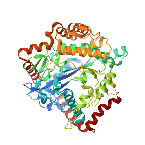 | |
UniProt | |||||
Find proteins for Q5NHS8 (Francisella tularensis subsp. tularensis (strain SCHU S4 / Schu 4)) Explore Q5NHS8 Go to UniProtKB: Q5NHS8 | |||||
Entity Groups | |||||
| Sequence Clusters | 30% Identity50% Identity70% Identity90% Identity95% Identity100% Identity | ||||
| UniProt Group | Q5NHS8 | ||||
Sequence AnnotationsExpand | |||||
| |||||
| Ligands 2 Unique | |||||
|---|---|---|---|---|---|
| ID | Chains | Name / Formula / InChI Key | 2D Diagram | 3D Interactions | |
| AMP Query on AMP | B [auth A] | ADENOSINE MONOPHOSPHATE C10 H14 N5 O7 P UDMBCSSLTHHNCD-KQYNXXCUSA-N |  | ||
| SO4 Query on SO4 | C [auth A] D [auth A] E [auth A] F [auth A] G [auth A] | SULFATE ION O4 S QAOWNCQODCNURD-UHFFFAOYSA-L |  | ||
| Length ( Å ) | Angle ( ˚ ) |
|---|---|
| a = 89.849 | α = 90 |
| b = 89.849 | β = 90 |
| c = 151.468 | γ = 120 |
| Software Name | Purpose |
|---|---|
| Blu-Ice | data collection |
| PHASER | phasing |
| REFMAC | refinement |
| HKL-3000 | data reduction |
| HKL-3000 | data scaling |
RCSB PDB (citation) is hosted by
RCSB PDB is a member of the

