3NYO

Crystal Structure of G Protein-Coupled Receptor Kinase 6 in Complex with AMP


Experimental Data Snapshot

  • Method: X-RAY DIFFRACTION
  • Resolution: 2.92 Å
  • R-Value Free: 0.253 
  • R-Value Work: 0.222 
  • R-Value Observed: 0.222 

wwPDB Validation   3D Report Full Report


Ligand Structure Quality Assessment 


This is version 1.2 of the entry. See complete history


Literature

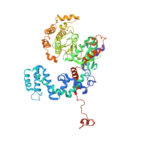
Molecular basis for activation of G protein-coupled receptor kinases.

Boguth, C.A.Singh, P.Huang, C.C.Tesmer, J.J.

(2010) EMBO J 29: 3249-3259

  • DOI: https://doi.org/10.1038/emboj.2010.206
  • Primary Citation of Related Structures:  
    3NYN, 3NYO

  • PubMed Abstract: 

    G protein-coupled receptor (GPCR) kinases (GRKs) selectively recognize and are allosterically regulated by activated GPCRs, but the molecular basis for this interaction is not understood. Herein, we report crystal structures of GRK6 in which regions known to be critical for receptor phosphorylation have coalesced to stabilize the kinase domain in a closed state and to form a likely receptor docking site. The crux of this docking site is an extended N-terminal helix that bridges the large and small lobes of the kinase domain and lies adjacent to a basic surface of the protein proposed to bind anionic phospholipids. Mutation of exposed, hydrophobic residues in the N-terminal helix selectively inhibits receptor, but not peptide phosphorylation, suggesting that these residues interact directly with GPCRs. Our structural and biochemical results thus provide an explanation for how receptor recognition, phospholipid binding, and kinase activation are intimately coupled in GRKs.


  • Organizational Affiliation

    Life Sciences Institute, The University of Michigan, Ann Arbor, MI, USA.


Macromolecules
Find similar proteins by:  (by identity cutoff)  |  3D Structure
Entity ID: 1
MoleculeChains Sequence LengthOrganismDetailsImage
G protein-coupled receptor kinase 6
A, B
576Homo sapiensMutation(s): 1 
Gene Names: GPRK6GRK6
EC: 2.7.11.16
UniProt & NIH Common Fund Data Resources
Find proteins for P43250 (Homo sapiens)
Explore P43250 
Go to UniProtKB:  P43250
PHAROS:  P43250
GTEx:  ENSG00000198055 
Entity Groups  
Sequence Clusters30% Identity50% Identity70% Identity90% Identity95% Identity100% Identity
UniProt GroupP43250
Sequence Annotations
Expand
  • Reference Sequence
Small Molecules
Ligands 3 Unique
IDChains Name / Formula / InChI Key2D Diagram3D Interactions
AMP
Query on AMP

Download Ideal Coordinates CCD File 
C [auth A],
M [auth B]
ADENOSINE MONOPHOSPHATE
C10 H14 N5 O7 P
UDMBCSSLTHHNCD-KQYNXXCUSA-N
SO4
Query on SO4

Download Ideal Coordinates CCD File 
D [auth A]
E [auth A]
F [auth A]
G [auth A]
H [auth A]
D [auth A],
E [auth A],
F [auth A],
G [auth A],
H [auth A],
J [auth A],
K [auth A],
L [auth A],
N [auth B],
O [auth B],
P [auth B],
Q [auth B],
S [auth B]
SULFATE ION
O4 S
QAOWNCQODCNURD-UHFFFAOYSA-L
BU3
Query on BU3

Download Ideal Coordinates CCD File 
I [auth A],
R [auth B]
(R,R)-2,3-BUTANEDIOL
C4 H10 O2
OWBTYPJTUOEWEK-QWWZWVQMSA-N
Experimental Data & Validation

Experimental Data

  • Method: X-RAY DIFFRACTION
  • Resolution: 2.92 Å
  • R-Value Free: 0.253 
  • R-Value Work: 0.222 
  • R-Value Observed: 0.222 
  • Space Group: P 61
Unit Cell:
Length ( Å )Angle ( ˚ )
a = 154.574α = 90
b = 154.574β = 90
c = 208.117γ = 120
Software Package:
Software NamePurpose
SCALEPACKdata scaling
PHASERphasing
REFMACrefinement
PDB_EXTRACTdata extraction
HKL-2000data reduction

Structure Validation

View Full Validation Report


Ligand Structure Quality Assessment 


Entry History 

Deposition Data

Revision History  (Full details and data files)

  • Version 1.0: 2010-09-22
    Type: Initial release
  • Version 1.1: 2011-07-13
    Changes: Version format compliance
  • Version 1.2: 2024-02-21
    Changes: Data collection, Database references, Derived calculations, Refinement description