3NY3

Structure of the ubr-box of UBR2 in complex with N-degron


Experimental Data Snapshot

  • Method: X-RAY DIFFRACTION
  • Resolution: 1.60 Å
  • R-Value Free: 0.210 
  • R-Value Work: 0.190 
  • R-Value Observed: 0.191 

wwPDB Validation   3D Report Full Report


This is version 1.2 of the entry. See complete history


Literature

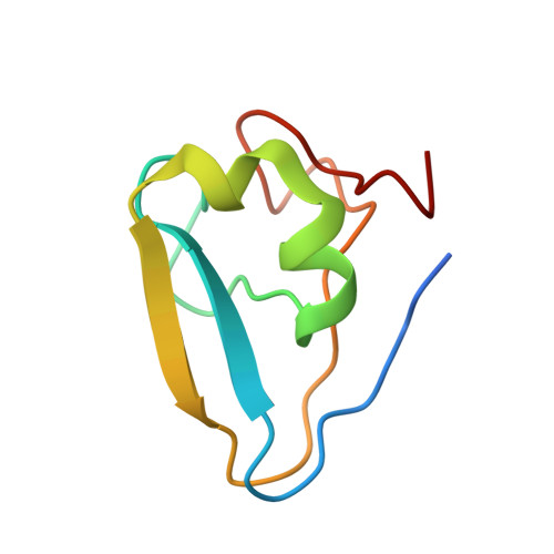
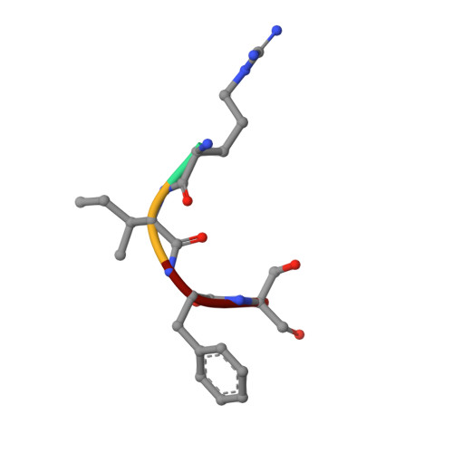
Structural basis of substrate recognition and specificity in the N-end rule pathway.

Matta-Camacho, E.Kozlov, G.Li, F.F.Gehring, K.

(2010) Nat Struct Mol Biol 17: 1182-1187

  • DOI: https://doi.org/10.1038/nsmb.1894
  • Primary Citation of Related Structures:  
    3NY1, 3NY2, 3NY3

  • PubMed Abstract: 

    The N-end rule links the half-life of a protein to the identity of its N-terminal residue. Destabilizing N-terminal residues are recognized by E3 ubiquitin ligases, termed N-recognins. A conserved structural domain called the UBR box is responsible for their specificity. Here we report the crystal structures of the UBR boxes of the human N-recognins UBR1 and UBR2, alone and in complex with an N-end rule peptide, Arg-Ile-Phe-Ser. These structures show that the UBR box adopts a previously undescribed fold stabilized through the binding of three zinc ions to form a binding pocket for type 1 N-degrons. NMR experiments reveal a preference for N-terminal arginine. Peptide binding is abrogated by N-terminal acetylation of the peptide or loss of the positive charge of the N-terminal residue. These results rationalize and refine the empirical rules for the classification of type 1 N-degrons. We also confirm that a missense mutation in UBR1 that is responsible for Johanson-Blizzard syndrome leads to UBR box unfolding and loss of function.


  • Organizational Affiliation

    Department of Biochemistry, McGill University, Montreal, Quebec, Canada.


Macromolecules
Find similar proteins by:  (by identity cutoff)  |  3D Structure
Entity ID: 1
MoleculeChains Sequence LengthOrganismDetailsImage
E3 ubiquitin-protein ligase UBR275Homo sapiensMutation(s): 0 
Gene Names: UBR2C6orf133KIAA0349
EC: 6.3.2.19
UniProt & NIH Common Fund Data Resources
Find proteins for Q8IWV8 (Homo sapiens)
Explore Q8IWV8 
Go to UniProtKB:  Q8IWV8
PHAROS:  Q8IWV8
GTEx:  ENSG00000024048 
Entity Groups  
Sequence Clusters30% Identity50% Identity70% Identity90% Identity95% Identity100% Identity
UniProt GroupQ8IWV8
Sequence Annotations
Expand
  • Reference Sequence

Find similar proteins by:  Sequence   |   3D Structure  

Entity ID: 2
MoleculeChains Sequence LengthOrganismDetailsImage
N-degron4N/AMutation(s): 0 
Sequence Annotations
Expand
  • Reference Sequence
Experimental Data & Validation

Experimental Data

  • Method: X-RAY DIFFRACTION
  • Resolution: 1.60 Å
  • R-Value Free: 0.210 
  • R-Value Work: 0.190 
  • R-Value Observed: 0.191 
  • Space Group: P 1 21 1
Unit Cell:
Length ( Å )Angle ( ˚ )
a = 29.123α = 90
b = 36.94β = 109.6
c = 29.824γ = 90
Software Package:
Software NamePurpose
CrystalCleardata collection
PHASERphasing
REFMACrefinement
HKL-2000data reduction
HKL-2000data scaling

Structure Validation

View Full Validation Report


Entry History 

Deposition Data

Revision History  (Full details and data files)

  • Version 1.0: 2010-08-11
    Type: Initial release
  • Version 1.1: 2011-07-13
    Changes: Version format compliance
  • Version 1.2: 2024-02-21
    Changes: Data collection, Database references, Derived calculations