


Polymer-Induced Heteronucleation for Protein Single Crystal Growth: Structural Elucidation of Bovine Liver Catalase and Concanavalin A Forms
Foroughi, L.M., Kang, Y.N., Matzger, A.J.(2011) Cryst Growth Des 11: 1294-1298
Experimental Data Snapshot
(2011) Cryst Growth Des 11: 1294-1298
Entity ID: 1 | |||||
|---|---|---|---|---|---|
| Molecule | Chains | Sequence Length | Organism | Details | Image |
| Catalase | 527 | Bos taurus | Mutation(s): 0 EC: 1.11.1.6 | 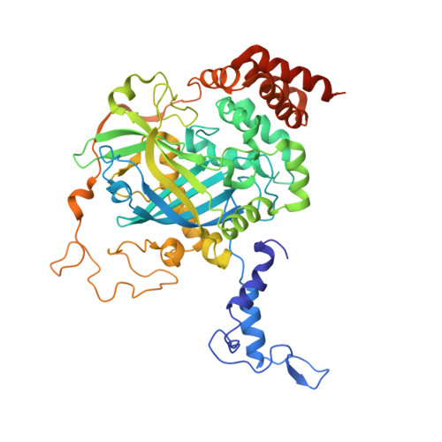 | |
UniProt | |||||
Find proteins for P00432 (Bos taurus) Explore P00432 Go to UniProtKB: P00432 | |||||
Entity Groups | |||||
| Sequence Clusters | 30% Identity50% Identity70% Identity90% Identity95% Identity100% Identity | ||||
| UniProt Group | P00432 | ||||
Sequence AnnotationsExpand | |||||
| |||||
| Ligands 2 Unique | |||||
|---|---|---|---|---|---|
| ID | Chains | Name / Formula / InChI Key | 2D Diagram | 3D Interactions | |
| NDP Query on NDP | F [auth A], H [auth B], J [auth C], L [auth D] | NADPH DIHYDRO-NICOTINAMIDE-ADENINE-DINUCLEOTIDE PHOSPHATE C21 H30 N7 O17 P3 ACFIXJIJDZMPPO-NNYOXOHSSA-N |  | ||
| HEM Query on HEM | E [auth A], G [auth B], I [auth C], K [auth D] | PROTOPORPHYRIN IX CONTAINING FE C34 H32 Fe N4 O4 KABFMIBPWCXCRK-RGGAHWMASA-L |  | ||
| Length ( Å ) | Angle ( ˚ ) |
|---|---|
| a = 68.652 | α = 90 |
| b = 173.744 | β = 90 |
| c = 186.325 | γ = 90 |
| Software Name | Purpose |
|---|---|
| HKL-2000 | data collection |
| PHASER | phasing |
| REFMAC | refinement |
| HKL-2000 | data reduction |
| HKL-2000 | data scaling |
RCSB PDB (citation) is hosted by
RCSB PDB is a member of the

