3NSZ

Human CK2 catalytic domain in complex with AMPPN


Experimental Data Snapshot

  • Method: X-RAY DIFFRACTION
  • Resolution: 1.30 Å
  • R-Value Free: 0.187 
  • R-Value Work: 0.168 
  • R-Value Observed: 0.169 

wwPDB Validation   3D Report Full Report


Ligand Structure Quality Assessment 


This is version 1.2 of the entry. See complete history


Literature

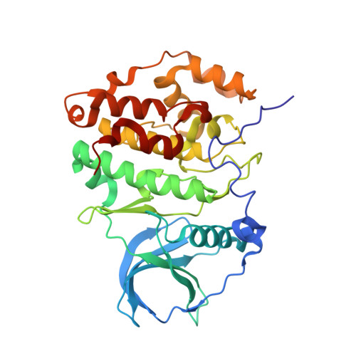
Structural basis of CX-4945 binding to human protein kinase CK2.

Ferguson, A.D.Sheth, P.R.Basso, A.D.Paliwal, S.Gray, K.Fischmann, T.O.Le, H.V.

(2011) FEBS Lett 585: 104-110

  • DOI: https://doi.org/10.1016/j.febslet.2010.11.019
  • Primary Citation of Related Structures:  
    3NGA, 3NSZ

  • PubMed Abstract: 

    Protein kinase CK2 (CK2), a constitutively active serine/threonine kinase, is involved in a variety of roles essential to the maintenance of cellular homeostasis. Elevated levels of CK2 expression results in the dysregulation of key signaling pathways that regulate transcription, and has been implicated in cancer. The adenosine-5'-triphosphate-competitive inhibitor CX-4945 has been reported to show broad spectrum anti-proliferative activity in multiple cancer cell lines. Although the enzymatic IC(50) of CX-4945 has been reported, the thermodynamics and structural basis of binding to CK2α remained elusive. Presented here are the crystal structures of human CK2α in complex with CX-4945 and adenylyl phosphoramidate at 2.7 and 1.3 Å, respectively. Biophysical analysis of CX-4945 binding is also described. This data provides the structural rationale for the design of more potent inhibitors against this emerging cancer target.


  • Organizational Affiliation

    Drug Design Department, Merck Research Laboratory, Kenilworth, NJ 07033, USA. andrew.ferguson@astrazeneca.com


Macromolecules
Find similar proteins by:  (by identity cutoff)  |  3D Structure
Entity ID: 1
MoleculeChains Sequence LengthOrganismDetailsImage
Casein kinase II subunit alpha330Homo sapiensMutation(s): 0 
Gene Names: CSNK2A1CK2A1
EC: 2.7.11.1
UniProt & NIH Common Fund Data Resources
Find proteins for P68400 (Homo sapiens)
Explore P68400 
Go to UniProtKB:  P68400
PHAROS:  P68400
GTEx:  ENSG00000101266 
Entity Groups  
Sequence Clusters30% Identity50% Identity70% Identity90% Identity95% Identity100% Identity
UniProt GroupP68400
Sequence Annotations
Expand
  • Reference Sequence
Experimental Data & Validation

Experimental Data

  • Method: X-RAY DIFFRACTION
  • Resolution: 1.30 Å
  • R-Value Free: 0.187 
  • R-Value Work: 0.168 
  • R-Value Observed: 0.169 
  • Space Group: P 21 21 21
Unit Cell:
Length ( Å )Angle ( ˚ )
a = 49.382α = 90
b = 62.026β = 90
c = 117.061γ = 90
Software Package:
Software NamePurpose
CrystalCleardata collection
MOLREPphasing
BUSTERrefinement
HKL-2000data reduction
HKL-2000data scaling

Structure Validation

View Full Validation Report


Ligand Structure Quality Assessment 


Entry History 

Deposition Data

Revision History  (Full details and data files)

  • Version 1.0: 2010-12-15
    Type: Initial release
  • Version 1.1: 2011-07-13
    Changes: Version format compliance
  • Version 1.2: 2023-12-27
    Changes: Data collection, Database references, Derived calculations