3NPD

Crystal structure of a putative secreted protein (PA3611) from PSEUDOMONAS AERUGINOSA at 1.60 A resolution


Experimental Data Snapshot

  • Method: X-RAY DIFFRACTION
  • Resolution: 1.60 Å
  • R-Value Free: 0.205 
  • R-Value Work: 0.160 
  • R-Value Observed: 0.162 

wwPDB Validation   3D Report Full Report


Ligand Structure Quality Assessment 


This is version 1.3 of the entry. See complete history


Literature

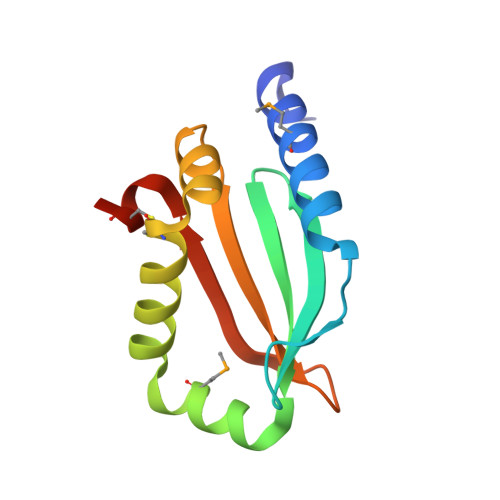
Crystal structure of a putative quorum sensing-regulated protein (PA3611) from the Pseudomonas-specific DUF4146 family.

Das, D.Chiu, H.J.Farr, C.L.Grant, J.C.Jaroszewski, L.Knuth, M.W.Miller, M.D.Tien, H.J.Elsliger, M.A.Deacon, A.M.Godzik, A.Lesley, S.A.Wilson, I.A.

(2014) Proteins 82: 1086-1092

  • DOI: https://doi.org/10.1002/prot.24455
  • Primary Citation of Related Structures:  
    3NPD

  • PubMed Abstract: 

    Pseudomonas aeruginosa is an opportunistic pathogen commonly found in humans and other organisms and is an important cause of infection especially in patients with compromised immune defense mechanisms. The PA3611 gene of P. aeruginosa PAO1 encodes a secreted protein of unknown function, which has been recently classified into a small Pseudomonas-specific protein family called DUF4146. As part of our effort to extend structural coverage of novel protein space and provide a structure-based functional insight into new protein families, we report the crystal structure of PA3611, the first structural representative of the DUF4146 protein family.


  • Organizational Affiliation

    Joint Center for Structural Genomics; Stanford Synchrotron Radiation Lightsource, SLAC National Accelerator Laboratory, Menlo Park, California.


Macromolecules
Find similar proteins by:  (by identity cutoff)  |  3D Structure
Entity ID: 1
MoleculeChains Sequence LengthOrganismDetailsImage
putative secreted protein118Pseudomonas aeruginosaMutation(s): 0 
Gene Names: PA3611
UniProt
Find proteins for Q9HY15 (Pseudomonas aeruginosa (strain ATCC 15692 / DSM 22644 / CIP 104116 / JCM 14847 / LMG 12228 / 1C / PRS 101 / PAO1))
Explore Q9HY15 
Go to UniProtKB:  Q9HY15
Entity Groups  
Sequence Clusters30% Identity50% Identity70% Identity90% Identity95% Identity100% Identity
UniProt GroupQ9HY15
Sequence Annotations
Expand
  • Reference Sequence
Small Molecules
Ligands 3 Unique
IDChains Name / Formula / InChI Key2D Diagram3D Interactions
CXS
Query on CXS

Download Ideal Coordinates CCD File 
D [auth A],
E [auth A],
F [auth A],
G [auth A]
3-CYCLOHEXYL-1-PROPYLSULFONIC ACID
C9 H19 N O3 S
PJWWRFATQTVXHA-UHFFFAOYSA-N
SO4
Query on SO4

Download Ideal Coordinates CCD File 
B [auth A],
C [auth A]
SULFATE ION
O4 S
QAOWNCQODCNURD-UHFFFAOYSA-L
EDO
Query on EDO

Download Ideal Coordinates CCD File 
H [auth A],
I [auth A]
1,2-ETHANEDIOL
C2 H6 O2
LYCAIKOWRPUZTN-UHFFFAOYSA-N
Modified Residues  1 Unique
IDChains TypeFormula2D DiagramParent
MSE
Query on MSE
A
L-PEPTIDE LINKINGC5 H11 N O2 SeMET
Experimental Data & Validation

Experimental Data

Unit Cell:
Length ( Å )Angle ( ˚ )
a = 34.989α = 90
b = 51.317β = 90
c = 56.898γ = 90
Software Package:
Software NamePurpose
SOLVEphasing
REFMACrefinement
XSCALEdata scaling
PDB_EXTRACTdata extraction
XDSdata reduction

Structure Validation

View Full Validation Report


Ligand Structure Quality Assessment 


Entry History 

Deposition Data

Revision History  (Full details and data files)

  • Version 1.0: 2010-07-28
    Type: Initial release
  • Version 1.1: 2011-07-13
    Changes: Version format compliance
  • Version 1.2: 2014-09-24
    Changes: Database references
  • Version 1.3: 2023-02-01
    Changes: Database references, Derived calculations