3NOG

Designed ankyrin repeat protein (DARPin) Binders to AcrB: Plasticity of the Interface


Experimental Data Snapshot

  • Method: X-RAY DIFFRACTION
  • Resolution: 3.34 Å
  • R-Value Free: 0.307 
  • R-Value Work: 0.257 
  • R-Value Observed: 0.258 

wwPDB Validation   3D Report Full Report


This is version 1.2 of the entry. See complete history


Literature

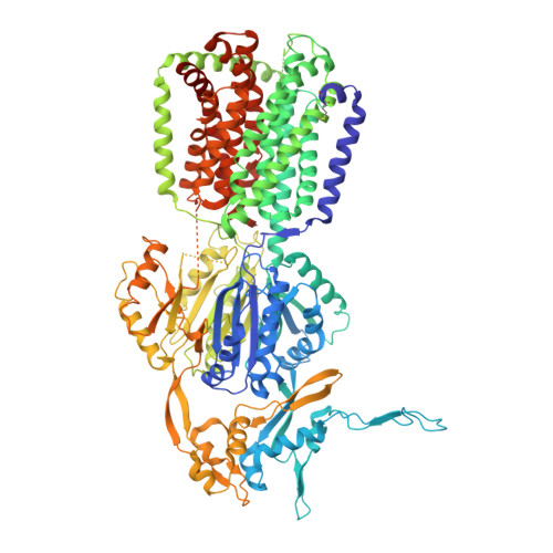
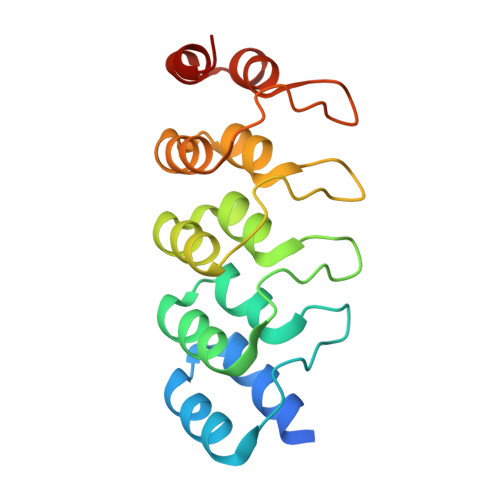
Designed ankyrin repeat protein binders for the crystallization of AcrB: Plasticity of the dominant interface

Monroe, N.Sennhauser, G.Seeger, M.A.Briand, C.Grutter, M.G.

(2011) J Struct Biol 174: 269-281

  • DOI: https://doi.org/10.1016/j.jsb.2011.01.014
  • Primary Citation of Related Structures:  
    3NOC, 3NOG

  • PubMed Abstract: 

    The formation of well-diffracting crystals is a major bottleneck in structural analysis of membrane proteins by X-ray crystallography. One approach to improve crystal quality is the use of DARPins as crystallization chaperones. Here, we present a detailed analysis of the interaction between DARPins and the integral membrane protein AcrB. We find that binders selected in vitro by ribosome display share a common epitope. The comparative analysis of three crystal structures of AcrB-DARPin complexes allowed us to study the plasticity of the interaction with this dominant binding site. Seemingly redundant AcrB-DARPin crystals show substantially different diffraction quality as a result of subtle differences in the binding geometry. This work exemplifies the importance to screen a number of crystallization chaperones to obtain optimal diffraction data. Crystallographic analysis is complemented by biophysical characterization of nine AcrB binders. We observe that small variations in the interface can lead to differing behavior of the DARPins with regards to affinity, stoichiometry of the complexes and specificity for their target.


  • Organizational Affiliation

    Department of Biochemistry, University of Zurich, Winterthurerstrasse 190, 8057 Zurich, Switzerland. nmonroe@bioc.uzh.ch


Macromolecules
Find similar proteins by:  (by identity cutoff)  |  3D Structure
Entity ID: 1
MoleculeChains Sequence LengthOrganismDetailsImage
Acriflavine resistance protein B
A, B, C
1,049Escherichia coli K-12Mutation(s): 0 
Gene Names: acrBacrEb0462JW0451
Membrane Entity: Yes 
UniProt
Find proteins for P31224 (Escherichia coli (strain K12))
Explore P31224 
Go to UniProtKB:  P31224
Entity Groups  
Sequence Clusters30% Identity50% Identity70% Identity90% Identity95% Identity100% Identity
UniProt GroupP31224
Sequence Annotations
Expand
  • Reference Sequence
Find similar proteins by:  (by identity cutoff)  |  3D Structure
Entity ID: 2
MoleculeChains Sequence LengthOrganismDetailsImage
Designed ankyrin repeat protein
D, E
169synthetic constructMutation(s): 0 
Entity Groups  
Sequence Clusters30% Identity50% Identity70% Identity90% Identity95% Identity100% Identity
Sequence Annotations
Expand
  • Reference Sequence
Small Molecules
Modified Residues  1 Unique
IDChains TypeFormula2D DiagramParent
FME
Query on FME
A, B, C
L-PEPTIDE LINKINGC6 H11 N O3 SMET
Experimental Data & Validation

Experimental Data

  • Method: X-RAY DIFFRACTION
  • Resolution: 3.34 Å
  • R-Value Free: 0.307 
  • R-Value Work: 0.257 
  • R-Value Observed: 0.258 
  • Space Group: P 21 21 21
Unit Cell:
Length ( Å )Angle ( ˚ )
a = 145.4α = 90
b = 158β = 90
c = 258.6γ = 90
Software Package:
Software NamePurpose
XSCALEdata scaling
PHASERphasing
PHENIXrefinement
PDB_EXTRACTdata extraction
XDSdata scaling
XDSdata reduction

Structure Validation

View Full Validation Report


Entry History 

Deposition Data

Revision History  (Full details and data files)

  • Version 1.0: 2011-05-18
    Type: Initial release
  • Version 1.1: 2011-07-13
    Changes: Version format compliance
  • Version 1.2: 2023-11-01
    Changes: Data collection, Database references, Derived calculations, Refinement description