


Crystal Structure of apo fumarate hydratase from Mycobacterium tuberculosis
Li, H., Swanson, S., Yu, M., Hung, L.-H., Sacchettini, J.S.To be published.
Experimental Data Snapshot
wwPDB Validation 3D Report Full Report

Entity ID: 1 | |||||
|---|---|---|---|---|---|
| Molecule | Chains | Sequence Length | Organism | Details | Image |
| Fumarate hydratase class II | 475 | Mycobacterium tuberculosis H37Rv | Mutation(s): 0 Gene Names: fum, fumC, MT1130, MTV017.51c, Rv1098c EC: 4.2.1.2 | 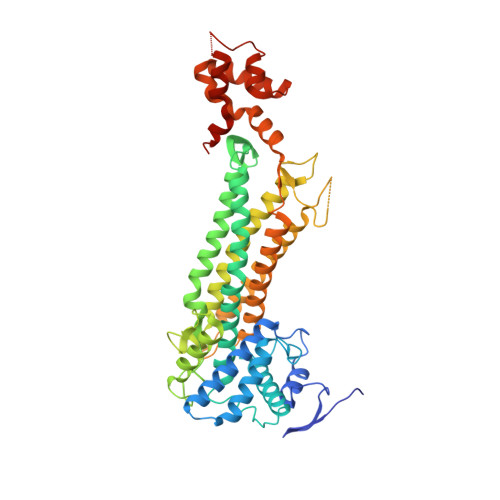 | |
UniProt | |||||
Find proteins for P9WN93 (Mycobacterium tuberculosis (strain ATCC 25618 / H37Rv)) Explore P9WN93 Go to UniProtKB: P9WN93 | |||||
Entity Groups | |||||
| Sequence Clusters | 30% Identity50% Identity70% Identity90% Identity95% Identity100% Identity | ||||
| UniProt Group | P9WN93 | ||||
Sequence AnnotationsExpand | |||||
| |||||
| Length ( Å ) | Angle ( ˚ ) |
|---|---|
| a = 271.224 | α = 90 |
| b = 96.555 | β = 102.99 |
| c = 89.889 | γ = 90 |
| Software Name | Purpose |
|---|---|
| MOLREP | phasing |
| PHENIX | refinement |
| HKL-2000 | data collection |
| DENZO | data reduction |
| SCALEPACK | data scaling |
RCSB PDB (citation) is hosted by
RCSB PDB is a member of the

