


Peptide-binding single chain Antibody fragment (SCFV) chaperones for protein co-crystallization
Pai, J., Culver, J.A., Drury, J.E., Lieberman, R.L., Maynard, J.A.To be published.
Experimental Data Snapshot
wwPDB Validation 3D Report Full Report

Entity ID: 1 | |||||
|---|---|---|---|---|---|
| Molecule | Chains | Sequence Length | Organism | Details | Image |
| Engineered scFv | 256 | Mus musculus | Mutation(s): 0 | 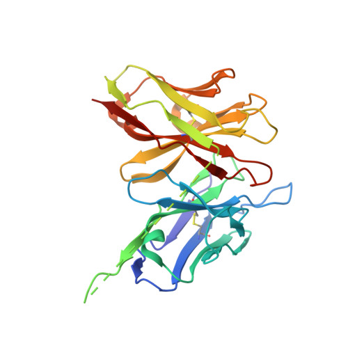 | |
Entity Groups | |||||
| Sequence Clusters | 30% Identity50% Identity70% Identity90% Identity95% Identity100% Identity | ||||
Sequence AnnotationsExpand | |||||
| |||||
| Length ( Å ) | Angle ( ˚ ) |
|---|---|
| a = 266.637 | α = 90 |
| b = 266.637 | β = 90 |
| c = 266.637 | γ = 90 |
| Software Name | Purpose |
|---|---|
| SCALA | data scaling |
| MOLREP | phasing |
| REFMAC | refinement |
| PDB_EXTRACT | data extraction |
| MAR345dtb | data collection |
| XSCALE | data scaling |
RCSB PDB (citation) is hosted by
RCSB PDB is a member of the

