3NHA

Nucleotide Binding Domain of Human ABCB6 (ADP Mg bound structure)


Experimental Data Snapshot

  • Method: X-RAY DIFFRACTION
  • Resolution: 2.10 Å
  • R-Value Free: 0.252 
  • R-Value Work: 0.184 
  • R-Value Observed: 0.187 

wwPDB Validation   3D Report Full Report


Ligand Structure Quality Assessment 


This is version 1.4 of the entry. See complete history


Literature

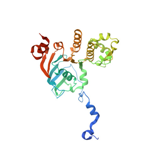
Structures of the nucleotide-binding domain of the human ABCB6 transporter and its complexes with nucleotides.

Haffke, M.Menzel, A.Carius, Y.Jahn, D.Heinz, D.W.

(2010) Acta Crystallogr D Biol Crystallogr 66: 979-987

  • DOI: https://doi.org/10.1107/S0907444910028593
  • Primary Citation of Related Structures:  
    3NH6, 3NH9, 3NHA, 3NHB

  • PubMed Abstract: 

    The human ATP-binding cassette (ABC) transporter ABCB6 is involved in haem-precursor transport across the mitochondrial membrane. The crystal structure of its nucleotide-binding domain (NBD) has been determined in the apo form and in complexes with ADP, with ADP and Mg(2+) and with ATP at high resolution. The overall structure is L-shaped and consists of two lobes, consistent with other reported NBD structures. Nucleotide binding is mediated by the highly conserved Tyr599 and the Walker A motif, and induces notable structural changes. Structural comparison with other structurally characterized NBDs and full-length ABC transporters gives the first insight into the possible catalytic mechanism of ABCB6 and the role of the N-terminal helix alpha(1) in full-length ABCB6.


  • Organizational Affiliation

    Helmholtz Zentrum für Infektionsforschung, Braunschweig, Germany.


Macromolecules
Find similar proteins by:  (by identity cutoff)  |  3D Structure
Entity ID: 1
MoleculeChains Sequence LengthOrganismDetailsImage
ATP-binding cassette sub-family B member 6, mitochondrial306Homo sapiensMutation(s): 0 
Gene Names: ABCB6MTABC3PRPUMAT
UniProt & NIH Common Fund Data Resources
Find proteins for Q9NP58 (Homo sapiens)
Explore Q9NP58 
Go to UniProtKB:  Q9NP58
PHAROS:  Q9NP58
GTEx:  ENSG00000115657 
Entity Groups  
Sequence Clusters30% Identity50% Identity70% Identity90% Identity95% Identity100% Identity
UniProt GroupQ9NP58
Sequence Annotations
Expand
  • Reference Sequence
Experimental Data & Validation

Experimental Data

  • Method: X-RAY DIFFRACTION
  • Resolution: 2.10 Å
  • R-Value Free: 0.252 
  • R-Value Work: 0.184 
  • R-Value Observed: 0.187 
  • Space Group: P 21 21 21
Unit Cell:
Length ( Å )Angle ( ˚ )
a = 56.272α = 90
b = 70.708β = 90
c = 71.113γ = 90
Software Package:
Software NamePurpose
XSCALEdata scaling
MOLREPphasing
REFMACrefinement
PDB_EXTRACTdata extraction
AUTOMARdata collection
XDSdata reduction

Structure Validation

View Full Validation Report


Ligand Structure Quality Assessment 


Entry History 

Deposition Data

Revision History  (Full details and data files)

  • Version 1.0: 2010-08-25
    Type: Initial release
  • Version 1.1: 2011-07-13
    Changes: Version format compliance
  • Version 1.2: 2012-03-21
    Changes: Database references
  • Version 1.3: 2017-09-27
    Changes: Data collection
  • Version 1.4: 2023-09-06
    Changes: Data collection, Database references, Derived calculations, Refinement description