3NG4

Ternary complex of peptidoglycan recognition protein (PGRP-S) with Maltose and N-Acetylglucosamine at 1.7 A Resolution


Experimental Data Snapshot

  • Method: X-RAY DIFFRACTION
  • Resolution: 1.73 Å
  • R-Value Free: 0.244 
  • R-Value Work: 0.200 
  • R-Value Observed: 0.203 

wwPDB Validation   3D Report Full Report


Ligand Structure Quality Assessment 


This is version 2.1 of the entry. See complete history


Literature

Multiligand specificity of pathogen-associated molecular pattern-binding site in peptidoglycan recognition protein

Sharma, P.Dube, D.Sinha, M.Mishra, B.Dey, S.Mal, G.Pathak, K.M.Kaur, P.Sharma, S.Singh, T.P.

(2011) J Biol Chem 286: 31723-31730

  • DOI: https://doi.org/10.1074/jbc.M111.264374
  • Primary Citation of Related Structures:  
    3NG4, 3NW3

  • PubMed Abstract: 

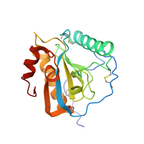
    The peptidoglycan recognition protein PGRP-S is an innate immunity molecule that specifically interacts with microbial peptidoglycans and other pathogen-associated molecular patterns. We report here two structures of the unique tetrameric camel PGRP-S (CPGRP-S) complexed with (i) muramyl dipeptide (MDP) at 2.5 Å resolution and (ii) GlcNAc and β-maltose at 1.7Å resolution. The binding studies carried out using surface plasmon resonance indicated that CPGRP-S binds to MDP with a dissociation constant of 10(-7) M, whereas the binding affinities for GlcNAc and β-maltose separately are in the range of 10(-4) M to 10(-5) M, whereas the dissociation constant for the mixture of GlcNAc and maltose was estimated to be 10(-6) M. The data from bacterial suspension culture experiments showed a significant inhibition of the growth of Staphylococcus aureus cells when CPGRP-S was added to culture medium. The ELISA experiment showed that the amount of MDP-induced production of TNF-α and IL-6 decreased considerably after the introduction of CPGRP-S. The crystal structure determinations of (i) a binary complex with MDP and (ii) a ternary complex with GlcNAc and β-maltose revealed that MDP, GlcNAc, and β-maltose bound to CPGRP-S in the ligand binding cleft, which is situated at the interface of molecules C and D of the homotetramer formed by four protein molecules A, B, C, and D. In the binary complex, the muramyl moiety of MDP is observed at the C-D interface, whereas the peptide chain protrudes into the center of tetramer. In the ternary complex, GlcNAc and β-maltose occupy distinct non-overlapping positions belonging to different subsites.


  • Organizational Affiliation

    Department of Biophysics, All India Institute of Medical Sciences, New Delhi, India 110029.


Macromolecules
Find similar proteins by:  (by identity cutoff)  |  3D Structure
Entity ID: 1
MoleculeChains Sequence LengthOrganismDetailsImage
Peptidoglycan recognition protein 1
A, B, C, D
171Camelus dromedariusMutation(s): 0 
UniProt
Find proteins for Q9GK12 (Camelus dromedarius)
Explore Q9GK12 
Go to UniProtKB:  Q9GK12
Entity Groups  
Sequence Clusters30% Identity50% Identity70% Identity90% Identity95% Identity100% Identity
UniProt GroupQ9GK12
Sequence Annotations
Expand
  • Reference Sequence
Oligosaccharides

Help

Entity ID: 2
MoleculeChains Length2D Diagram Glycosylation3D Interactions
alpha-D-glucopyranose-(1-4)-alpha-D-glucopyranose
E
2N/A
Glycosylation Resources
GlyTouCan:  G07411ON
GlyCosmos:  G07411ON
Small Molecules
Biologically Interesting Molecules (External Reference) 1 Unique
Experimental Data & Validation

Experimental Data

  • Method: X-RAY DIFFRACTION
  • Resolution: 1.73 Å
  • R-Value Free: 0.244 
  • R-Value Work: 0.200 
  • R-Value Observed: 0.203 
  • Space Group: I 2 2 2
Unit Cell:
Length ( Å )Angle ( ˚ )
a = 87.128α = 90
b = 100.784β = 90
c = 161.85γ = 90
Software Package:
Software NamePurpose
HKL-2000data collection
AMoREphasing
REFMACrefinement
HKL-2000data reduction
HKL-2000data scaling

Structure Validation

View Full Validation Report


Ligand Structure Quality Assessment 


Entry History 

Deposition Data

Revision History  (Full details and data files)

  • Version 1.0: 2010-07-14
    Type: Initial release
  • Version 1.1: 2011-07-13
    Changes: Version format compliance
  • Version 1.2: 2012-06-13
    Changes: Database references
  • Version 2.0: 2020-07-29
    Type: Remediation
    Reason: Carbohydrate remediation
    Changes: Atomic model, Data collection, Derived calculations, Non-polymer description, Structure summary
  • Version 2.1: 2023-11-01
    Changes: Data collection, Database references, Refinement description, Structure summary