3NEO

Wild type human transthyretin (TTR) complexed with GC-24 (TTRwt:GC-24)


Experimental Data Snapshot

  • Method: X-RAY DIFFRACTION
  • Resolution: 2.00 Å
  • R-Value Free: 0.220 
  • R-Value Work: 0.183 
  • R-Value Observed: 0.185 

wwPDB Validation   3D Report Full Report


Ligand Structure Quality Assessment 


This is version 1.2 of the entry. See complete history


Literature

The binding of synthetic triiodo l-thyronine analogs to human transthyretin: molecular basis of cooperative and non-cooperative ligand recognition.

Trivella, D.B.Sairre, M.I.Foguel, D.Lima, L.M.Polikarpov, I.

(2011) J Struct Biol 173: 323-332

  • DOI: https://doi.org/10.1016/j.jsb.2010.10.003
  • Primary Citation of Related Structures:  
    3NEE, 3NEO, 3NES, 3NEX

  • PubMed Abstract: 

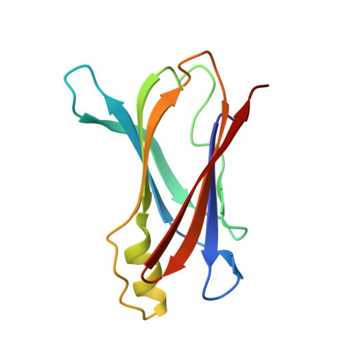
    Transthyretin (TTR) is a tetrameric β-sheet-rich transporter protein directly involved in human amyloid diseases. Several classes of small molecules can bind to TTR delaying its amyloid fibril formation, thus being promising drug candidates to treat TTR amyloidoses. In the present study, we characterized the interactions of the synthetic triiodo L-thyronine analogs and thyroid hormone nuclear receptor TRβ-selective agonists GC-1 and GC-24 with the wild type and V30M variant of human transthyretin (TTR). To achieve this aim, we conducted in vitro TTR acid-mediated aggregation and isothermal titration calorimetry experiments and determined the TTR:GC-1 and TTR:GC-24 crystal structures. Our data indicate that both GC-1 and GC-24 bind to TTR in a non-cooperative manner and are good inhibitors of TTR aggregation, with dissociation constants for both hormone binding sites (HBS) in the low micromolar range. Analysis of the crystal structures of TTRwt:GC-1(24) complexes and their comparison with the TTRwt X-ray structure bound to its natural ligand thyroxine (T4) suggests, at the molecular level, the basis for the cooperative process displayed by T4 and the non-cooperative process provoked by both GC-1 and GC-24 during binding to TTR.


  • Organizational Affiliation

    Instituto de Física de São Carlos-Universidade de São Paulo, São Carlos, SP, Brazil.


Macromolecules
Find similar proteins by:  (by identity cutoff)  |  3D Structure
Entity ID: 1
MoleculeChains Sequence LengthOrganismDetailsImage
Transthyretin
A, B
116Homo sapiensMutation(s): 0 
Gene Names: PALBTTR
UniProt & NIH Common Fund Data Resources
Find proteins for P02766 (Homo sapiens)
Explore P02766 
Go to UniProtKB:  P02766
PHAROS:  P02766
GTEx:  ENSG00000118271 
Entity Groups  
Sequence Clusters30% Identity50% Identity70% Identity90% Identity95% Identity100% Identity
UniProt GroupP02766
Sequence Annotations
Expand
  • Reference Sequence
Small Molecules
Ligands 1 Unique
IDChains Name / Formula / InChI Key2D Diagram3D Interactions
G24
Query on G24

Download Ideal Coordinates CCD File 
C [auth A],
D [auth B]
[4-(3-BENZYL-4-HYDROXYBENZYL)-3,5-DIMETHYLPHENOXY]ACETIC ACID
C24 H24 O4
JYHIGYLGYNCMGI-UHFFFAOYSA-N
Binding Affinity Annotations 
IDSourceBinding Affinity
G24 PDBBind:  3NEO Kd: 1200 (nM) from 1 assay(s)
Experimental Data & Validation

Experimental Data

  • Method: X-RAY DIFFRACTION
  • Resolution: 2.00 Å
  • R-Value Free: 0.220 
  • R-Value Work: 0.183 
  • R-Value Observed: 0.185 
  • Space Group: P 21 21 2
Unit Cell:
Length ( Å )Angle ( ˚ )
a = 42.77α = 90
b = 84.91β = 90
c = 64.44γ = 90
Software Package:
Software NamePurpose
SCALAdata scaling
PHASERphasing
REFMACrefinement
PDB_EXTRACTdata extraction
MAR345dtbdata collection
MOSFLMdata reduction

Structure Validation

View Full Validation Report


Ligand Structure Quality Assessment 


Entry History 

Deposition Data

Revision History  (Full details and data files)

  • Version 1.0: 2010-11-24
    Type: Initial release
  • Version 1.1: 2011-07-13
    Changes: Version format compliance
  • Version 1.2: 2024-02-21
    Changes: Data collection, Database references, Derived calculations