3NEA

Crystal Structure of Peptidyl-tRNA hydrolase from Francisella tularensis


Experimental Data Snapshot

  • Method: X-RAY DIFFRACTION
  • Resolution: 2.25 Å
  • R-Value Free: 0.248 
  • R-Value Work: 0.190 
  • R-Value Observed: 0.193 

wwPDB Validation   3D Report Full Report


This is version 1.3 of the entry. See complete history


Literature

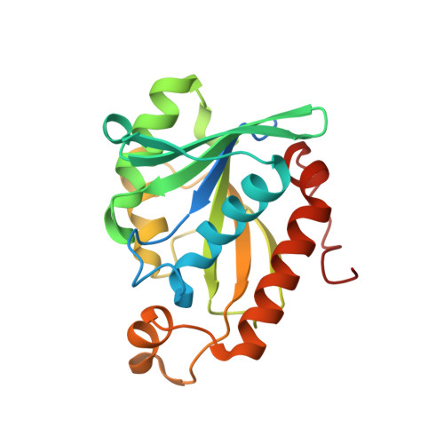
Structure of Francisella tularensis peptidyl-tRNA hydrolase.

Clarke, T.E.Romanov, V.Lam, R.Gothe, S.A.Peddi, S.R.Razumova, E.B.Lipman, R.S.Branstrom, A.A.Chirgadze, N.Y.

(2011) Acta Crystallogr Sect F Struct Biol Cryst Commun 67: 446-449

  • DOI: https://doi.org/10.1107/S174430911100515X
  • Primary Citation of Related Structures:  
    3NEA

  • PubMed Abstract: 

    The rational design of novel antibiotics for bacteria involves the identification of inhibitors for enzymes involved in essential biochemical pathways in cells. In this study, the cloning, expression, purification, crystallization and structure of the enzyme peptidyl-tRNA hydrolase from Francisella tularensis, the causative agent of tularemia, was performed. The structure of F. tularensis peptidyl-tRNA hydrolase is comparable to those of other bacterial peptidyl-tRNA hydrolases, with most residues in the active site conserved amongst the family. The resultant reagents, structural data and analyses provide essential information for the structure-based design of novel inhibitors for this class of proteins.


  • Organizational Affiliation

    Division of Cancer Genomics and Proteomics, Ontario Cancer Institute, University Health Network, MBRC 5th Floor, 200 Elizabeth Street, Toronto, Ontario M5G 2C4, Canada.


Macromolecules
Find similar proteins by:  (by identity cutoff)  |  3D Structure
Entity ID: 1
MoleculeChains Sequence LengthOrganismDetailsImage
Peptidyl-tRNA hydrolase207Francisella tularensis subsp. tularensisMutation(s): 0 
Gene Names: FTT_0680cpth
EC: 3.1.1.29
UniProt
Find proteins for Q5NGZ6 (Francisella tularensis subsp. tularensis (strain SCHU S4 / Schu 4))
Explore Q5NGZ6 
Go to UniProtKB:  Q5NGZ6
Entity Groups  
Sequence Clusters30% Identity50% Identity70% Identity90% Identity95% Identity100% Identity
UniProt GroupQ5NGZ6
Sequence Annotations
Expand
  • Reference Sequence
Experimental Data & Validation

Experimental Data

  • Method: X-RAY DIFFRACTION
  • Resolution: 2.25 Å
  • R-Value Free: 0.248 
  • R-Value Work: 0.190 
  • R-Value Observed: 0.193 
  • Space Group: P 21 21 2
Unit Cell:
Length ( Å )Angle ( ˚ )
a = 60.683α = 90
b = 93.043β = 90
c = 33.027γ = 90
Software Package:
Software NamePurpose
DENZOdata reduction
SCALEPACKdata scaling
MOLREPphasing
REFMACrefinement
PDB_EXTRACTdata extraction
CrystalCleardata collection
HKL-2000data reduction
HKL-2000data scaling

Structure Validation

View Full Validation Report


Entry History 

Deposition Data

Revision History  (Full details and data files)

  • Version 1.0: 2011-06-15
    Type: Initial release
  • Version 1.1: 2011-07-13
    Changes: Version format compliance
  • Version 1.2: 2017-11-08
    Changes: Refinement description
  • Version 1.3: 2023-09-06
    Changes: Data collection, Database references, Derived calculations, Refinement description