3NC0

Crystal structure of the HIV-1 Rev NES-CRM1-RanGTP nuclear export complex (crystal II)


Experimental Data Snapshot

  • Method: X-RAY DIFFRACTION
  • Resolution: 2.90 Å
  • R-Value Free: 0.295 
  • R-Value Work: 0.242 
  • R-Value Observed: 0.245 

wwPDB Validation   3D Report Full Report


Ligand Structure Quality Assessment 


This is version 1.3 of the entry. See complete history


Literature

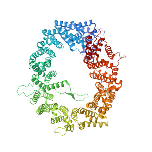
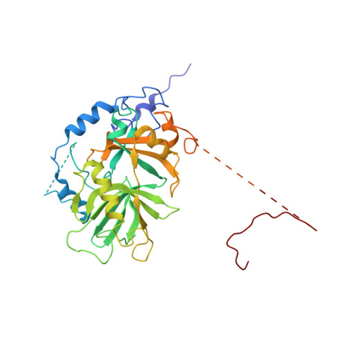
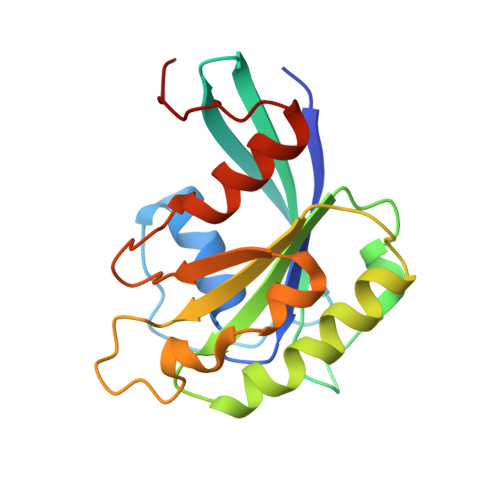
NES consensus redefined by structures of PKI-type and Rev-type nuclear export signals bound to CRM1.

Guttler, T.Madl, T.Neumann, P.Deichsel, D.Corsini, L.Monecke, T.Ficner, R.Sattler, M.Gorlich, D.

(2010) Nat Struct Mol Biol 17: 1367-1376

  • DOI: https://doi.org/10.1038/nsmb.1931
  • Primary Citation of Related Structures:  
    2L1L, 3NBY, 3NBZ, 3NC0, 3NC1

  • PubMed Abstract: 

    Classic nuclear export signals (NESs) confer CRM1-dependent nuclear export. Here we present crystal structures of the RanGTP-CRM1 complex alone and bound to the prototypic PKI or HIV-1 Rev NESs. These NESs differ markedly in the spacing of their key hydrophobic (Φ) residues, yet CRM1 recognizes them with the same rigid set of five Φ pockets. The different Φ spacings are compensated for by different conformations of the bound NESs: in the case of PKI, an α-helical conformation, and in the case of Rev, an extended conformation with a critical proline docking into a Φ pocket. NMR analyses of CRM1-bound and CRM1-free PKI NES suggest that CRM1 selects NES conformers that pre-exist in solution. Our data lead to a new structure-based NES consensus, and explain why NESs differ in their affinities for CRM1 and why supraphysiological NESs bind the exportin so tightly.


  • Organizational Affiliation

    Max-Planck-Institut für Biophysikalische Chemie, Göttingen, Germany.


Macromolecules
Find similar proteins by:  (by identity cutoff)  |  3D Structure
Entity ID: 1
MoleculeChains Sequence LengthOrganismDetailsImage
Exportin-1
A, D
1,073Mus musculusMutation(s): 0 
Gene Names: Crm1Xpo1Xpo1 (GeneID: 103573)
UniProt & NIH Common Fund Data Resources
Find proteins for Q6P5F9 (Mus musculus)
Explore Q6P5F9 
Go to UniProtKB:  Q6P5F9
IMPC:  MGI:2144013
Entity Groups  
Sequence Clusters30% Identity50% Identity70% Identity90% Identity95% Identity100% Identity
UniProt GroupQ6P5F9
Sequence Annotations
Expand
  • Reference Sequence
Find similar proteins by:  (by identity cutoff)  |  3D Structure
Entity ID: 2
MoleculeChains Sequence LengthOrganismDetailsImage
Snurportin-1
B, E
362Homo sapiensMutation(s): 0 
Gene Names: SNUPN (GeneID: 10073)RNUT1SNUPNSPN1
UniProt & NIH Common Fund Data Resources
Find proteins for O95149 (Homo sapiens)
Explore O95149 
Go to UniProtKB:  O95149
GTEx:  ENSG00000169371 
Entity Groups  
Sequence Clusters30% Identity50% Identity70% Identity90% Identity95% Identity100% Identity
UniProt GroupO95149
Sequence Annotations
Expand
  • Reference Sequence
Find similar proteins by:  (by identity cutoff)  |  3D Structure
Entity ID: 3
MoleculeChains Sequence LengthOrganismDetailsImage
GTP-binding nuclear protein Ran
C, F
176Homo sapiensMutation(s): 1 
Gene Names: ARA24OK/SW-cl.81RANRAN (GeneID: 5901)
UniProt & NIH Common Fund Data Resources
Find proteins for P62826 (Homo sapiens)
Explore P62826 
Go to UniProtKB:  P62826
PHAROS:  P62826
GTEx:  ENSG00000132341 
Entity Groups  
Sequence Clusters30% Identity50% Identity70% Identity90% Identity95% Identity100% Identity
UniProt GroupP62826
Sequence Annotations
Expand
  • Reference Sequence
Small Molecules
Ligands 5 Unique
IDChains Name / Formula / InChI Key2D Diagram3D Interactions
GTP
Query on GTP

Download Ideal Coordinates CCD File 
EA [auth F],
Q [auth C]
GUANOSINE-5'-TRIPHOSPHATE
C10 H16 N5 O14 P3
XKMLYUALXHKNFT-UUOKFMHZSA-N
PEG
Query on PEG

Download Ideal Coordinates CCD File 
AA [auth D]
GA [auth L]
J [auth A]
K [auth A]
L [auth A]
AA [auth D],
GA [auth L],
J [auth A],
K [auth A],
L [auth A],
M [auth A],
T [auth C],
V [auth D],
W [auth D],
X [auth D],
Y [auth D],
Z [auth D]
DI(HYDROXYETHYL)ETHER
C4 H10 O3
MTHSVFCYNBDYFN-UHFFFAOYSA-N
IPH
Query on IPH

Download Ideal Coordinates CCD File 
O [auth B]PHENOL
C6 H6 O
ISWSIDIOOBJBQZ-UHFFFAOYSA-N
GOL
Query on GOL

Download Ideal Coordinates CCD File 
BA [auth D]
CA [auth E]
DA [auth E]
G [auth A]
H [auth A]
BA [auth D],
CA [auth E],
DA [auth E],
G [auth A],
H [auth A],
I [auth A],
N [auth A],
P [auth B],
S [auth C],
U [auth D]
GLYCEROL
C3 H8 O3
PEDCQBHIVMGVHV-UHFFFAOYSA-N
MG
Query on MG

Download Ideal Coordinates CCD File 
FA [auth F],
R [auth C]
MAGNESIUM ION
Mg
JLVVSXFLKOJNIY-UHFFFAOYSA-N
Experimental Data & Validation

Experimental Data

  • Method: X-RAY DIFFRACTION
  • Resolution: 2.90 Å
  • R-Value Free: 0.295 
  • R-Value Work: 0.242 
  • R-Value Observed: 0.245 
  • Space Group: P 1 21 1
Unit Cell:
Length ( Å )Angle ( ˚ )
a = 73.262α = 90
b = 225.895β = 100.75
c = 163.984γ = 90
Software Package:
Software NamePurpose
XSCALEdata processing
PHENIXrefinement
PDB_EXTRACTdata extraction
MAR345dtbdata collection
XDSdata reduction
XSCALEdata scaling
CNSphasing

Structure Validation

View Full Validation Report


Ligand Structure Quality Assessment 


Entry History 

Deposition Data

Revision History  (Full details and data files)

  • Version 1.0: 2010-10-27
    Type: Initial release
  • Version 1.1: 2011-07-13
    Changes: Version format compliance
  • Version 1.2: 2021-10-06
    Changes: Database references, Derived calculations
  • Version 1.3: 2023-09-06
    Changes: Data collection, Refinement description