3NB3

The host outer membrane proteins OmpA and OmpC are packed at specific sites in the Shigella phage Sf6 virion as structural components


Experimental Data Snapshot

  • Method: ELECTRON MICROSCOPY
  • Resolution: 19.0 Å
  • Aggregation State: PARTICLE 
  • Reconstruction Method: SINGLE PARTICLE 

wwPDB Validation   3D Report Full Report


This is version 1.5 of the entry. See complete history


Literature

The host outer membrane proteins OmpA and OmpC are associated with the Shigella phage Sf6 virion.

Zhao, H.Sequeira, R.D.Galeva, N.A.Tang, L.

(2011) Virology 409: 319-327

  • DOI: https://doi.org/10.1016/j.virol.2010.10.030
  • Primary Citation of Related Structures:  
    3NB3

  • PubMed Abstract: 

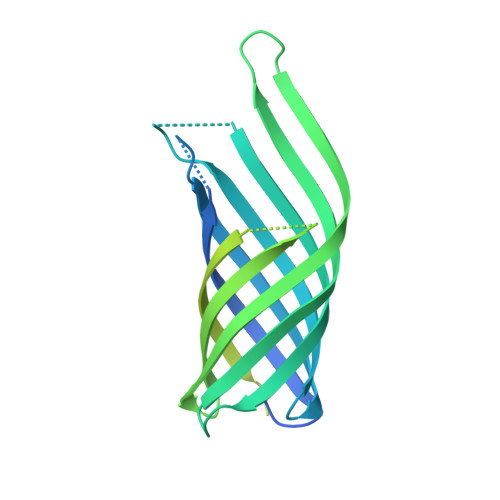
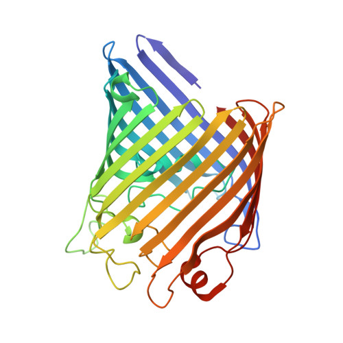
    Assembly of dsDNA bacteriophage is a precisely programmed process. Potential roles of host cell components in phage assembly haven't been well understood. It was previously reported that two unidentified proteins were present in bacteriophage Sf6 virion (Casjens et al, 2004, J.Mol.Biol. 339, 379-394, Fig. 2A). Using tandem mass spectrometry, we have identified the two proteins as outer membrane proteins (OMPs) OmpA and OmpC from its host Shigella flexneri. The transmission electron cryo-microscopy structure of Sf6 shows significant density at specific sites at the phage capsid inner surface. This density fit well with the characteristic beta-barrel domains of OMPs, thus may be due to the two host proteins. Locations of this density suggest a role in Sf6 morphogenesis reminiscent of phage-encoded cementing proteins. These data indicate a new, OMP-related phage:host linkage, adding to previous knowledge that some lambdoid bacteriophage genomes contain OmpC-like genes that express phage-encoded porins in the lysogenic state.


  • Organizational Affiliation

    Department of Molecular Biosciences, University of Kansas, 1200 Sunnyside Avenue, Lawrence, KS 66045, USA. zhaohy@ku.edu


Macromolecules
Find similar proteins by:  (by identity cutoff)  |  3D Structure
Entity ID: 1
MoleculeChains Sequence LengthOrganismDetailsImage
Outer membrane protein A
A, B, C
346Escherichia coli K-12Mutation(s): 0 
UniProt
Find proteins for P0A910 (Escherichia coli (strain K12))
Explore P0A910 
Go to UniProtKB:  P0A910
Entity Groups  
Sequence Clusters30% Identity50% Identity70% Identity90% Identity95% Identity100% Identity
UniProt GroupP0A910
Sequence Annotations
Expand
  • Reference Sequence
Find similar proteins by:  (by identity cutoff)  |  3D Structure
Entity ID: 2
MoleculeChains Sequence LengthOrganismDetailsImage
Outer membrane protein C346Escherichia coli K-12Mutation(s): 0 
UniProt
Find proteins for P06996 (Escherichia coli (strain K12))
Explore P06996 
Go to UniProtKB:  P06996
Entity Groups  
Sequence Clusters30% Identity50% Identity70% Identity90% Identity95% Identity100% Identity
UniProt GroupP06996
Sequence Annotations
Expand
  • Reference Sequence
Experimental Data & Validation

Experimental Data

  • Method: ELECTRON MICROSCOPY
  • Resolution: 19.0 Å
  • Aggregation State: PARTICLE 
  • Reconstruction Method: SINGLE PARTICLE 

Structure Validation

View Full Validation Report


Entry History 

Deposition Data

Revision History  (Full details and data files)

  • Version 1.0: 2011-02-16
    Type: Initial release
  • Version 1.1: 2011-07-13
    Changes: Version format compliance
  • Version 1.2: 2012-04-25
    Changes: Atomic model, Other, Source and taxonomy
  • Version 1.3: 2016-11-09
    Changes: Other
  • Version 1.4: 2019-12-11
    Changes: Data collection, Structure summary
  • Version 1.5: 2024-02-21
    Changes: Data collection, Database references, Derived calculations, Refinement description