Genetic and pharmacological inhibition of PDK1 in cancer cells: characterization of a selective allosteric kinase inhibitor.
Nagashima, K., Shumway, S.D., Sathyanarayanan, S., Chen, A.H., Dolinski, B., Xu, Y., Keilhack, H., Nguyen, T., Wiznerowicz, M., Li, L., Lutterbach, B.A., Chi, A., Paweletz, C., Allison, T., Yan, Y., Munshi, S.K., Klippel, A., Kraus, M., Bobkova, E.V., Deshmukh, S., Xu, Z., Mueller, U., Szewczak, A.A., Pan, B.S., Richon, V., Pollock, R., Blume-Jensen, P., Northrup, A., Andersen, J.N.(2011) J Biol Chem 286: 6433-6448
- PubMed: 21118801
- DOI: https://doi.org/10.1074/jbc.M110.156463
- Primary Citation of Related Structures:
3NAX - PubMed Abstract:
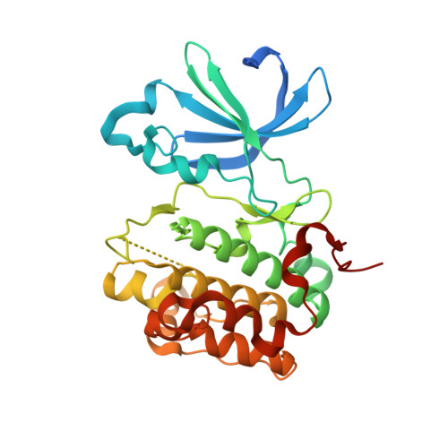
Phosphoinositide-dependent kinase 1 (PDK1) is a critical activator of multiple prosurvival and oncogenic protein kinases and has garnered considerable interest as an oncology drug target. Despite progress characterizing PDK1 as a therapeutic target, pharmacological support is lacking due to the prevalence of nonspecific inhibitors. Here, we benchmark literature and newly developed inhibitors and conduct parallel genetic and pharmacological queries into PDK1 function in cancer cells. Through kinase selectivity profiling and x-ray crystallographic studies, we identify an exquisitely selective PDK1 inhibitor (compound 7) that uniquely binds to the inactive kinase conformation (DFG-out). In contrast to compounds 1-5, which are classical ATP-competitive kinase inhibitors (DFG-in), compound 7 specifically inhibits cellular PDK1 T-loop phosphorylation (Ser-241), supporting its unique binding mode. Interfering with PDK1 activity has minimal antiproliferative effect on cells growing as plastic-attached monolayer cultures (i.e. standard tissue culture conditions) despite reduced phosphorylation of AKT, RSK, and S6RP. However, selective PDK1 inhibition impairs anchorage-independent growth, invasion, and cancer cell migration. Compound 7 inhibits colony formation in a subset of cancer cell lines (four of 10) and primary xenograft tumor lines (nine of 57). RNAi-mediated knockdown corroborates the PDK1 dependence in cell lines and identifies candidate biomarkers of drug response. In summary, our profiling studies define a uniquely selective and cell-potent PDK1 inhibitor, and the convergence of genetic and pharmacological phenotypes supports a role of PDK1 in tumorigenesis in the context of three-dimensional in vitro culture systems.
Organizational Affiliation:
Merck Research Laboratories, Boston, Massachusetts 02115, USA.