


The crystal structure of beta-phosphoglucomutase from Bacillus subtilis
Zhang, Z., Burley, S.K., Swaminathan, S.To be published.
Experimental Data Snapshot
wwPDB Validation 3D Report Full Report

Entity ID: 1 | |||||
|---|---|---|---|---|---|
| Molecule | Chains | Sequence Length | Organism | Details | Image |
| beta-phosphoglucomutase | 233 | Bacillus subtilis | Mutation(s): 0 Gene Names: BSU34550, yvdM EC: 5.4.2.6 | 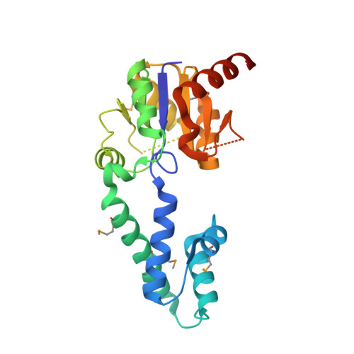 | |
UniProt | |||||
Find proteins for O06995 (Bacillus subtilis (strain 168)) Explore O06995 Go to UniProtKB: O06995 | |||||
Entity Groups | |||||
| Sequence Clusters | 30% Identity50% Identity70% Identity90% Identity95% Identity100% Identity | ||||
| UniProt Group | O06995 | ||||
Sequence AnnotationsExpand | |||||
| |||||
| Modified Residues 1 Unique | |||||
|---|---|---|---|---|---|
| ID | Chains | Type | Formula | 2D Diagram | Parent |
| MSE Query on MSE | A, B | L-PEPTIDE LINKING | C5 H11 N O2 Se |  | MET |
| Length ( Å ) | Angle ( ˚ ) |
|---|---|
| a = 67.043 | α = 90 |
| b = 78.171 | β = 90 |
| c = 92.067 | γ = 90 |
| Software Name | Purpose |
|---|---|
| CBASS | data collection |
| SOLVE | phasing |
| PHENIX | refinement |
| MOSFLM | data reduction |
| SCALA | data scaling |
RCSB PDB (citation) is hosted by
RCSB PDB is a member of the

