Biochemical and Structural Analysis of an Eis Family Aminoglycoside Acetyltransferase from Bacillus anthracis.
Green, K.D., Biswas, T., Chang, C., Wu, R., Chen, W., Janes, B.K., Chalupska, D., Gornicki, P., Hanna, P.C., Tsodikov, O.V., Joachimiak, A., Garneau-Tsodikova, S.(2015) Biochemistry 54: 3197-3206
- PubMed: 25928210
- DOI: https://doi.org/10.1021/acs.biochem.5b00244
- Primary Citation of Related Structures:
3N7Z - PubMed Abstract:
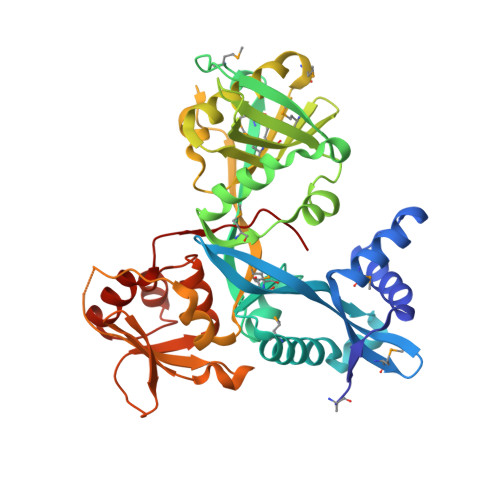
Proteins from the enhanced intracellular survival (Eis) family are versatile acetyltransferases that acetylate amines at multiple positions of several aminoglycosides (AGs). Their upregulation confers drug resistance. Homologues of Eis are present in diverse bacteria, including many pathogens. Eis from Mycobacterium tuberculosis (Eis_Mtb) has been well characterized. In this study, we explored the AG specificity and catalytic efficiency of the Eis family protein from Bacillus anthracis (Eis_Ban). Kinetic analysis of specificity and catalytic efficiency of acetylation of six AGs indicates that Eis_Ban displays significant differences from Eis_Mtb in both substrate binding and catalytic efficiency. The number of acetylated amines was also different for several AGs, indicating a distinct regiospecificity of Eis_Ban. Furthermore, most recently identified inhibitors of Eis_Mtb did not inhibit Eis_Ban, underscoring the differences between these two enzymes. To explain these differences, we determined an Eis_Ban crystal structure. The comparison of the crystal structures of Eis_Ban and Eis_Mtb demonstrates that critical residues lining their respective substrate binding pockets differ substantially, explaining their distinct specificities. Our results suggest that acetyltransferases of the Eis family evolved divergently to garner distinct specificities while conserving catalytic efficiency, possibly to counter distinct chemical challenges. The unique specificity features of these enzymes can be utilized as tools for developing AGs with novel modifications and help guide specific AG treatments to avoid Eis-mediated resistance.
Organizational Affiliation:
⊥Department of Pharmaceutical Sciences, University of Kentucky, Lexington, Kentucky 40536-0596, United States.