A structural hinge in eukaryotic MutY homologues mediates catalytic activity and Rad9-Rad1-Hus1 checkpoint complex interactions.
Luncsford, P.J., Chang, D.Y., Shi, G., Bernstein, J., Madabushi, A., Patterson, D.N., Lu, A.L., Toth, E.A.(2010) J Mol Biol 403: 351-370
- PubMed: 20816984
- DOI: https://doi.org/10.1016/j.jmb.2010.08.045
- Primary Citation of Related Structures:
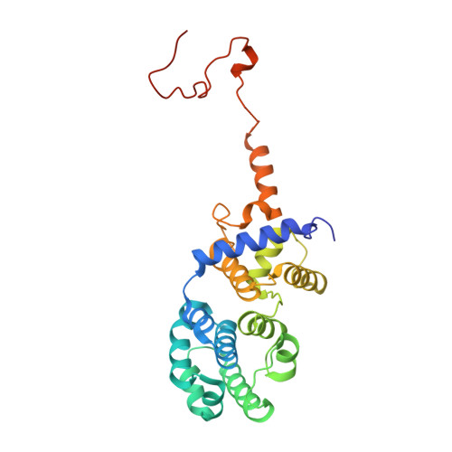
3N5N - PubMed Abstract:
The DNA glycosylase MutY homologue (MYH or MUTYH) removes adenines misincorporated opposite 8-oxoguanine as part of the base excision repair pathway. Importantly, defects in human MYH (hMYH) activity cause the inherited colorectal cancer syndrome MYH-associated polyposis. A key feature of MYH activity is its coordination with cell cycle checkpoint via interaction with the Rad9-Rad1-Hus1 (9-1-1) complex. The 9-1-1 complex facilitates cell cycle checkpoint activity and coordinates this activity with ongoing DNA repair. The interdomain connector (IDC, residues 295-350) between the catalytic domain and the 8-oxoguanine recognition domain of hMYH is a critical element that maintains interactions with the 9-1-1 complex. We report the first crystal structure of a eukaryotic MutY protein, a fragment of hMYH (residues 65-350) that consists of the catalytic domain and the IDC. Our structure reveals that the IDC adopts a stabilized conformation projecting away from the catalytic domain to form a docking scaffold for 9-1-1. We further examined the role of the IDC using Schizosaccharomyces pombe MYH as model system. In vitro studies of S. pombe MYH identified residues I261 and E262 of the IDC (equivalent to V315 and E316 of the hMYH IDC) as critical for maintaining the MYH/9-1-1 interaction. We determined that the eukaryotic IDC is also required for DNA damage selection and robust enzymatic activity. Our studies also provide the first evidence that disruption of the MYH/9-1-1 interaction diminishes the repair of oxidative DNA damage in vivo. Thus, preserving the MYH/9-1-1 interaction contributes significantly to minimizing the mutagenic potential of oxidative DNA damage.
Organizational Affiliation:
Department of Biochemistry and Molecular Biology, University of Maryland School of Medicine, 108 North Greene Street, Baltimore, MD 21201, USA.