3N3E

Zebrafish AlphaA crystallin


Experimental Data Snapshot

  • Method: X-RAY DIFFRACTION
  • Resolution: 1.75 Å
  • R-Value Free: 0.248 
  • R-Value Work: 0.205 
  • R-Value Observed: 0.207 

wwPDB Validation   3D Report Full Report


This is version 1.3 of the entry. See complete history


Literature

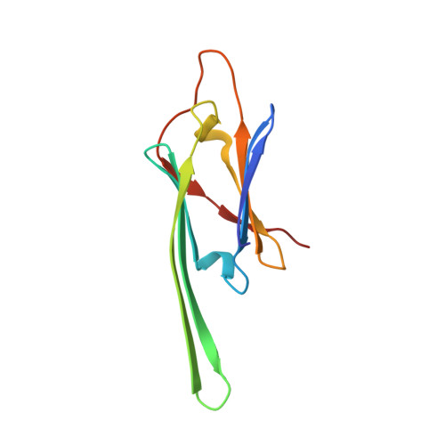
Non-3D domain swapped crystal structure of truncated zebrafish alphaA crystallin.

Laganowsky, A.Eisenberg, D.

(2010) Protein Sci 19: 1978-1984

  • DOI: https://doi.org/10.1002/pro.471
  • Primary Citation of Related Structures:  
    3N3E

  • PubMed Abstract: 

    In previous work on truncated alpha crystallins (Laganowsky et al., Protein Sci 2010; 19:1031-1043), we determined crystal structures of the alpha crystallin core, a seven beta-stranded immunoglobulin-like domain, with its conserved C-terminal extension. These extensions swap into neighboring cores forming oligomeric assemblies. The extension is palindromic in sequence, binding in either of two directions. Here, we report the crystal structure of a truncated alphaA crystallin (AAC) from zebrafish (Danio rerio) revealing C-terminal extensions in a non three-dimensional (3D) domain swapped, "closed" state. The extension is quasi-palindromic, bound within its own zebrafish core domain, lying in the opposite direction to that of bovine AAC, which is bound within an adjacent core domain (Laganowsky et al., Protein Sci 2010; 19:1031-1043). Our findings establish that the C-terminal extension of alpha crystallin proteins can be either 3D domain swapped or non-3D domain swapped. This duality provides another molecular mechanism for alpha crystallin proteins to maintain the polydispersity that is crucial for eye lens transparency.


  • Organizational Affiliation

    UCLA-DOE, Institute for Genomics and Proteomics, Los Angeles, California 90095-1570, USA.


Macromolecules
Find similar proteins by:  (by identity cutoff)  |  3D Structure
Entity ID: 1
MoleculeChains Sequence LengthOrganismDetailsImage
Alpha A crystallin
A, B
106Danio rerioMutation(s): 0 
Gene Names: cryaa
UniProt
Find proteins for Q8UUZ6 (Danio rerio)
Explore Q8UUZ6 
Go to UniProtKB:  Q8UUZ6
Entity Groups  
Sequence Clusters30% Identity50% Identity70% Identity90% Identity95% Identity100% Identity
UniProt GroupQ8UUZ6
Sequence Annotations
Expand
  • Reference Sequence
Experimental Data & Validation

Experimental Data

  • Method: X-RAY DIFFRACTION
  • Resolution: 1.75 Å
  • R-Value Free: 0.248 
  • R-Value Work: 0.205 
  • R-Value Observed: 0.207 
  • Space Group: P 43 21 2
Unit Cell:
Length ( Å )Angle ( ˚ )
a = 33.297α = 90
b = 33.297β = 90
c = 354.337γ = 90
Software Package:
Software NamePurpose
DENZOdata reduction
SCALEPACKdata scaling
PHASERphasing
PHENIXrefinement
PDB_EXTRACTdata extraction
ADSCdata collection

Structure Validation

View Full Validation Report


Entry History 

Deposition Data

Revision History  (Full details and data files)

  • Version 1.0: 2010-08-11
    Type: Initial release
  • Version 1.1: 2011-07-13
    Changes: Version format compliance
  • Version 1.2: 2017-11-08
    Changes: Refinement description
  • Version 1.3: 2023-09-06
    Changes: Data collection, Database references, Refinement description