3N0A

Crystal structure of auxilin (40-400)


Experimental Data Snapshot

  • Method: X-RAY DIFFRACTION
  • Resolution: 2.20 Å
  • R-Value Free: 0.200 
  • R-Value Work: 0.172 
  • R-Value Observed: 0.173 

wwPDB Validation   3D Report Full Report


This is version 1.2 of the entry. See complete history


Literature

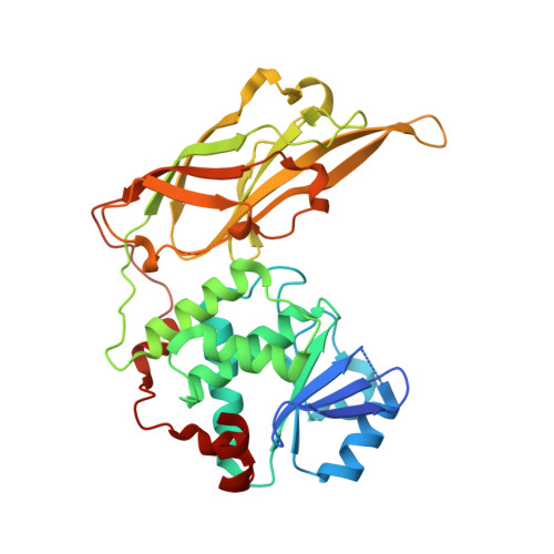
Structure of the PTEN-like Region of Auxilin, a Detector of Clathrin-Coated Vesicle Budding.

Guan, R.Han, D.Harrison, S.C.Kirchhausen, T.

(2010) Structure 18: 1191-1198

  • DOI: https://doi.org/10.1016/j.str.2010.06.016
  • Primary Citation of Related Structures:  
    3N0A

  • PubMed Abstract: 

    Auxilin, a J-domain containing protein, recruits the Hsc70 uncoating ATPase to newly budded clathrin-coated vesicles. The timing of auxilin arrival determines that uncoating will commence only after the clathrin lattice has fully assembled and after membrane fission is complete. Auxilin has a region resembling PTEN, a PI3P phosphatase. We have determined the crystal structure of this region of bovine auxilin 1; it indeed resembles PTEN closely. A change in the structure of the P loop accounts for the lack of phosphatase activity. Inclusion of phosphatidylinositol phosphates substantially enhances liposome binding by wild-type auxilin, but not by various mutants bearing changes in loops of the C2 domain. Nearly all these mutations also prevent recruitment of auxilin to newly budded coated vesicles. We propose a specific geometry for auxilin association with a membrane bilayer and discuss implications of this model for the mechanism by which auxilin detects separation of a vesicle from its parent membrane.


  • Organizational Affiliation

    Department of Cell Biology, Harvard Medical School, Boston, MA 02115, USA.


Macromolecules
Find similar proteins by:  (by identity cutoff)  |  3D Structure
Entity ID: 1
MoleculeChains Sequence LengthOrganismDetailsImage
Tyrosine-protein phosphatase auxilin361Bos taurusMutation(s): 0 
Gene Names: DNAJC6
EC: 3.1.3.48
UniProt
Find proteins for Q27974 (Bos taurus)
Explore Q27974 
Go to UniProtKB:  Q27974
Entity Groups  
Sequence Clusters30% Identity50% Identity70% Identity90% Identity95% Identity100% Identity
UniProt GroupQ27974
Sequence Annotations
Expand
  • Reference Sequence
Experimental Data & Validation

Experimental Data

  • Method: X-RAY DIFFRACTION
  • Resolution: 2.20 Å
  • R-Value Free: 0.200 
  • R-Value Work: 0.172 
  • R-Value Observed: 0.173 
  • Space Group: P 32 2 1
Unit Cell:
Length ( Å )Angle ( ˚ )
a = 137.925α = 90
b = 137.925β = 90
c = 51.327γ = 120
Software Package:
Software NamePurpose
ADSCdata collection
PHASERphasing
PHENIXrefinement
HKL-2000data reduction
SCALEPACKdata scaling

Structure Validation

View Full Validation Report


Entry History 

Deposition Data

Revision History  (Full details and data files)

  • Version 1.0: 2010-09-22
    Type: Initial release
  • Version 1.1: 2011-07-13
    Changes: Version format compliance
  • Version 1.2: 2023-09-06
    Changes: Data collection, Database references, Derived calculations, Refinement description