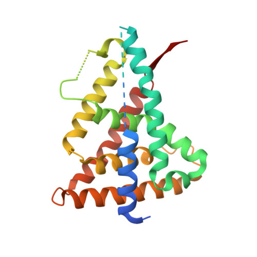
3N00

Crystal Structure of a deletion mutant of human Reverba ligand binding domain bound with an NCoR ID1 peptide determined to 2.60A


Experimental Data Snapshot

  • Method: X-RAY DIFFRACTION
  • Resolution: 2.60 Å
  • R-Value Free: 0.266 
  • R-Value Work: 0.199 
  • R-Value Observed: 0.200 

wwPDB Validation   3D Report Full Report


This is version 1.3 of the entry. See complete history


Literature

Structure of Rev-erbalpha bound to N-CoR reveals a unique mechanism of nuclear receptor-co-repressor interaction.

Phelan, C.A.Gampe, R.T.Lambert, M.H.Parks, D.J.Montana, V.Bynum, J.Broderick, T.M.Hu, X.Williams, S.P.Nolte, R.T.Lazar, M.A.

(2010) Nat Struct Mol Biol 17: 808-814

  • DOI: https://doi.org/10.1038/nsmb.1860
  • Primary Citation of Related Structures:  
    3N00

  • PubMed Abstract: 

    Repression of gene transcription by the nuclear receptor Rev-erbalpha plays an integral role in the core molecular circadian clock. We report the crystal structure of a nuclear receptor-co-repressor (N-CoR) interaction domain 1 (ID1) peptide bound to truncated human Rev-erbalpha ligand-binding domain (LBD). The ID1 peptide forms an unprecedented antiparallel beta-sheet with Rev-erbalpha, as well as an alpha-helix similar to that seen in nuclear receptor ID2 crystal structures but out of register by four residues. Comparison with the structure of Rev-erbbeta bound to heme indicates that ID1 peptide and heme induce substantially different conformational changes in the LBD. Although heme is involved in Rev-erb repression, the structure suggests that Rev-erbalpha could also mediate repression via ID1 binding in the absence of heme. The previously uncharacterized secondary structure induced by ID1 peptide binding advances our understanding of nuclear receptor-co-repressor interactions.


  • Organizational Affiliation

    Division of Endocrinology, Diabetes and Metabolism, Departments of Medicine and Genetics, The Institute for Diabetes, Obesity and Metabolism, University of Pennsylvania School of Medicine, Philadelphia, Pennsylvania, USA.


Macromolecules
Find similar proteins by:  (by identity cutoff)  |  3D Structure
Entity ID: 1
MoleculeChains Sequence LengthOrganismDetailsImage
Rev-erbA-alpha245Homo sapiensMutation(s): 0 
Gene Names: NR1D1EAR1HREVTHRAL
UniProt & NIH Common Fund Data Resources
Find proteins for P20393 (Homo sapiens)
Explore P20393 
Go to UniProtKB:  P20393
PHAROS:  P20393
GTEx:  ENSG00000126368 
Entity Groups  
Sequence Clusters30% Identity50% Identity70% Identity90% Identity95% Identity100% Identity
UniProt GroupP20393
Sequence Annotations
Expand
  • Reference Sequence

Find similar proteins by:  Sequence   |   3D Structure  

Entity ID: 2
MoleculeChains Sequence LengthOrganismDetailsImage
Nuclear receptor corepressor 121Homo sapiensMutation(s): 0 
UniProt & NIH Common Fund Data Resources
Find proteins for O75376 (Homo sapiens)
Explore O75376 
Go to UniProtKB:  O75376
PHAROS:  O75376
GTEx:  ENSG00000141027 
Entity Groups  
Sequence Clusters30% Identity50% Identity70% Identity90% Identity95% Identity100% Identity
UniProt GroupO75376
Sequence Annotations
Expand
  • Reference Sequence
Experimental Data & Validation

Experimental Data

  • Method: X-RAY DIFFRACTION
  • Resolution: 2.60 Å
  • R-Value Free: 0.266 
  • R-Value Work: 0.199 
  • R-Value Observed: 0.200 
  • Space Group: H 3 2
Unit Cell:
Length ( Å )Angle ( ˚ )
a = 112.55α = 90
b = 112.55β = 90
c = 103.833γ = 120
Software Package:
Software NamePurpose
JDirectordata collection
AMoREphasing
REFMACrefinement
HKL-2000data reduction
HKL-2000data scaling

Structure Validation

View Full Validation Report


Entry History 

Deposition Data

Revision History  (Full details and data files)

  • Version 1.0: 2010-06-30
    Type: Initial release
  • Version 1.1: 2011-07-13
    Changes: Version format compliance
  • Version 1.2: 2017-08-16
    Changes: Refinement description, Source and taxonomy
  • Version 1.3: 2023-09-06
    Changes: Data collection, Database references, Refinement description