3MOF

The structure of rat cytosolic PEPCK mutant A467G in complex with oxalate and GTP


Experimental Data Snapshot

  • Method: X-RAY DIFFRACTION
  • Resolution: 1.75 Å
  • R-Value Free: 0.228 
  • R-Value Work: 0.179 
  • R-Value Observed: 0.182 

wwPDB Validation   3D Report Full Report


Ligand Structure Quality Assessment 


This is version 1.4 of the entry. See complete history


Literature

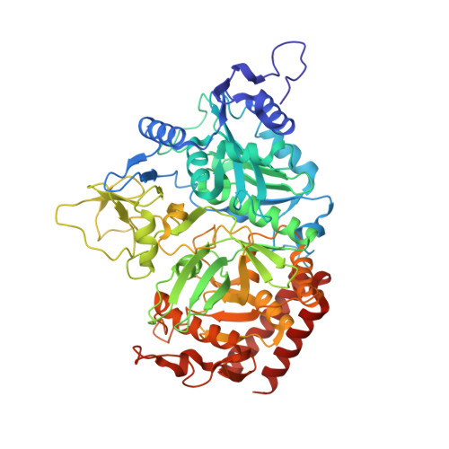
Increasing the conformational entropy of the Omega-loop lid domain in phosphoenolpyruvate carboxykinase impairs catalysis and decreases catalytic fidelity .

Johnson, T.A.Holyoak, T.

(2010) Biochemistry 49: 5176-5187

  • DOI: https://doi.org/10.1021/bi100399e
  • Primary Citation of Related Structures:  
    3MOE, 3MOF, 3MOH

  • PubMed Abstract: 

    Many studies have shown that the dynamic motions of individual protein segments can play an important role in enzyme function. Recent structural studies of the gluconeogenic enzyme phosphoenolpyruvate carboxykinase (PEPCK) demonstrate that PEPCK contains a 10-residue Omega-loop domain that acts as an active site lid. On the basis of these structural studies, we have previously proposed a model for the mechanism of PEPCK catalysis in which the conformation of this mobile lid domain is energetically coupled to ligand binding, resulting in the closed conformation of the lid, necessary for correct substrate positioning, becoming more energetically favorable as ligands associate with the enzyme. Here we test this model by introducing a point mutation (A467G) into the center of the Omega-loop lid that is designed to increase the entropic penalty for lid closure. Structural and kinetic characterization of this mutant enzyme demonstrates that the mutation has decreased the favorability of the enzyme adapting the closed lid conformation. As a consequence of this shift in the equilibrium defining the conformation of the active site lid, the enzyme's ability to stabilize the reaction intermediate is weakened, resulting in catalytic defect. This stabilization is initially surprising, as the lid domain makes no direct contacts with the enolate intermediate formed during the reaction. Furthermore, during the conversion of OAA to PEP, the destabilization of the lid-closed conformation results in the reaction becoming decoupled as the enolate intermediate is protonated rather than phosphorylated, resulting in the formation of pyruvate. Taken together, the structural and kinetic characterization of A467G-PEPCK supports our model of the role of the active site lid in catalytic function and demonstrates that the shift in the lowest-energy conformation between open and closed lid states is a function of the free energy available to the enzyme through ligand binding and the entropic penalty for ordering of the 10-residue Omega-loop lid domain.


  • Organizational Affiliation

    Department of Biochemistry and Molecular Biology, The University of Kansas Medical Center, Kansas City, Kansas 66160, USA.


Macromolecules
Find similar proteins by:  (by identity cutoff)  |  3D Structure
Entity ID: 1
MoleculeChains Sequence LengthOrganismDetailsImage
Phosphoenolpyruvate carboxykinase, cytosolic [GTP]
A, B
624Rattus norvegicusMutation(s): 1 
Gene Names: Pck1
EC: 4.1.1.32
UniProt
Find proteins for P07379 (Rattus norvegicus)
Explore P07379 
Go to UniProtKB:  P07379
Entity Groups  
Sequence Clusters30% Identity50% Identity70% Identity90% Identity95% Identity100% Identity
UniProt GroupP07379
Sequence Annotations
Expand
  • Reference Sequence
Small Molecules
Ligands 6 Unique
IDChains Name / Formula / InChI Key2D Diagram3D Interactions
GTP
Query on GTP

Download Ideal Coordinates CCD File 
C [auth A],
M [auth B]
GUANOSINE-5'-TRIPHOSPHATE
C10 H16 N5 O14 P3
XKMLYUALXHKNFT-UUOKFMHZSA-N
1PE
Query on 1PE

Download Ideal Coordinates CCD File 
H [auth A],
I [auth A],
R [auth B]
PENTAETHYLENE GLYCOL
C10 H22 O6
JLFNLZLINWHATN-UHFFFAOYSA-N
OXL
Query on OXL

Download Ideal Coordinates CCD File 
F [auth A],
Q [auth B]
OXALATE ION
C2 O4
MUBZPKHOEPUJKR-UHFFFAOYSA-L
EDO
Query on EDO

Download Ideal Coordinates CCD File 
J [auth A]
K [auth A]
L [auth A]
S [auth B]
T [auth B]
J [auth A],
K [auth A],
L [auth A],
S [auth B],
T [auth B],
U [auth B]
1,2-ETHANEDIOL
C2 H6 O2
LYCAIKOWRPUZTN-UHFFFAOYSA-N
MN
Query on MN

Download Ideal Coordinates CCD File 
D [auth A],
E [auth A],
N [auth B],
O [auth B]
MANGANESE (II) ION
Mn
WAEMQWOKJMHJLA-UHFFFAOYSA-N
NA
Query on NA

Download Ideal Coordinates CCD File 
G [auth A],
P [auth B]
SODIUM ION
Na
FKNQFGJONOIPTF-UHFFFAOYSA-N
Binding Affinity Annotations 
IDSourceBinding Affinity
OXL PDBBind:  3MOF Ki: 8.60e+5 (nM) from 1 assay(s)
Binding MOAD:  3MOF Ki: 8.60e+5 (nM) from 1 assay(s)
Experimental Data & Validation

Experimental Data

  • Method: X-RAY DIFFRACTION
  • Resolution: 1.75 Å
  • R-Value Free: 0.228 
  • R-Value Work: 0.179 
  • R-Value Observed: 0.182 
  • Space Group: P 1 21 1
Unit Cell:
Length ( Å )Angle ( ˚ )
a = 61.983α = 90
b = 119.601β = 106.87
c = 87.068γ = 90
Software Package:
Software NamePurpose
DENZOdata reduction
SCALEPACKdata scaling
MOLREPphasing
REFMACrefinement
PDB_EXTRACTdata extraction
Blu-Icedata collection
HKL-2000data reduction
HKL-2000data scaling

Structure Validation

View Full Validation Report


Ligand Structure Quality Assessment 


Entry History 

Deposition Data

Revision History  (Full details and data files)

  • Version 1.0: 2010-06-02
    Type: Initial release
  • Version 1.1: 2011-07-13
    Changes: Advisory, Version format compliance
  • Version 1.2: 2017-11-08
    Changes: Refinement description
  • Version 1.3: 2021-10-06
    Changes: Database references, Derived calculations
  • Version 1.4: 2024-02-21
    Changes: Data collection