3MNG

wild type human PrxV with DTT bound as a competitive inhibitor


Experimental Data Snapshot

  • Method: X-RAY DIFFRACTION
  • Resolution: 1.45 Å
  • R-Value Free: 0.136 
  • R-Value Work: 0.113 
  • R-Value Observed: 0.114 

wwPDB Validation   3D Report Full Report


Ligand Structure Quality Assessment 


This is version 1.2 of the entry. See complete history


Literature

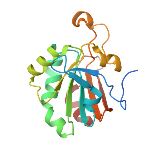
Structural Evidence that Peroxiredoxin Catalytic Power Is Based on Transition-State Stabilization.

Hall, A.Parsonage, D.Poole, L.B.Karplus, P.A.

(2010) J Mol Biol 402: 194-209

  • DOI: https://doi.org/10.1016/j.jmb.2010.07.022
  • Primary Citation of Related Structures:  
    3MNG

  • PubMed Abstract: 

    Peroxiredoxins (Prxs) are important peroxidases associated with both antioxidant protection and redox signaling. They use a conserved Cys residue to reduce peroxide substrates. The Prxs have a remarkably high catalytic efficiency that makes them a dominant player in cell-wide peroxide reduction, but the origins of their high activity have been mysterious. We present here a novel structure of human PrxV at 1.45 A resolution that has a dithiothreitol bound in the active site with its diol moiety mimicking the two oxygens of a peroxide substrate. This suggests diols and similar di-oxygen compounds as a novel class of competitive inhibitors for the Prxs. Common features of this and other structures containing peroxide, peroxide-mimicking ligands, or peroxide-mimicking water molecules reveal hydrogen bonding and steric factors that promote its high reactivity by creating an oxygen track along which the peroxide oxygens move as the reaction proceeds. Key insights include how the active-site microenvironment activates both the peroxidatic cysteine side chain and the peroxide substrate and how it is exquisitely well suited to stabilize the transition state of the in-line S(N)2 substitution reaction that is peroxidation.


  • Organizational Affiliation

    Department of Biochemistry and Biophysics, Oregon State University, Corvallis, OR 97331, USA.


Macromolecules
Find similar proteins by:  (by identity cutoff)  |  3D Structure
Entity ID: 1
MoleculeChains Sequence LengthOrganismDetailsImage
Peroxiredoxin-5, mitochondrial173Homo sapiensMutation(s): 0 
Gene Names: ACR1aoeb166arc1pmp20PRDX5SBBI10
EC: 1.11.1.15
UniProt & NIH Common Fund Data Resources
Find proteins for P30044 (Homo sapiens)
Explore P30044 
Go to UniProtKB:  P30044
PHAROS:  P30044
GTEx:  ENSG00000126432 
Entity Groups  
Sequence Clusters30% Identity50% Identity70% Identity90% Identity95% Identity100% Identity
UniProt GroupP30044
Sequence Annotations
Expand
  • Reference Sequence
Small Molecules
Ligands 3 Unique
IDChains Name / Formula / InChI Key2D Diagram3D Interactions
D1D
Query on D1D

Download Ideal Coordinates CCD File 
B [auth A](4S,5S)-1,2-DITHIANE-4,5-DIOL
C4 H8 O2 S2
YPGMOWHXEQDBBV-QWWZWVQMSA-N
GOL
Query on GOL

Download Ideal Coordinates CCD File 
K [auth A],
L [auth A]
GLYCEROL
C3 H8 O3
PEDCQBHIVMGVHV-UHFFFAOYSA-N
BR
Query on BR

Download Ideal Coordinates CCD File 
C [auth A]
D [auth A]
E [auth A]
F [auth A]
G [auth A]
C [auth A],
D [auth A],
E [auth A],
F [auth A],
G [auth A],
H [auth A],
I [auth A],
J [auth A]
BROMIDE ION
Br
CPELXLSAUQHCOX-UHFFFAOYSA-M
Experimental Data & Validation

Experimental Data

  • Method: X-RAY DIFFRACTION
  • Resolution: 1.45 Å
  • R-Value Free: 0.136 
  • R-Value Work: 0.113 
  • R-Value Observed: 0.114 
  • Space Group: P 41 21 2
Unit Cell:
Length ( Å )Angle ( ˚ )
a = 67.313α = 90
b = 67.313β = 90
c = 123.867γ = 90
Software Package:
Software NamePurpose
REFMACrefinement
MOSFLMdata reduction
SCALAdata scaling
REFMACphasing

Structure Validation

View Full Validation Report


Ligand Structure Quality Assessment 


Entry History 

Deposition Data

Revision History  (Full details and data files)

  • Version 1.0: 2010-08-04
    Type: Initial release
  • Version 1.1: 2011-07-13
    Changes: Version format compliance
  • Version 1.2: 2023-09-06
    Changes: Data collection, Database references, Derived calculations, Refinement description