3MMV

Structures of actin-bound WH2 domains of Spire and the implication for filament nucleation


Experimental Data Snapshot

  • Method: X-RAY DIFFRACTION
  • Resolution: 2.80 Å
  • R-Value Free: 0.330 
  • R-Value Work: 0.241 
  • R-Value Observed: 0.245 

wwPDB Validation   3D Report Full Report


Ligand Structure Quality Assessment 


This is version 1.4 of the entry. See complete history


Literature

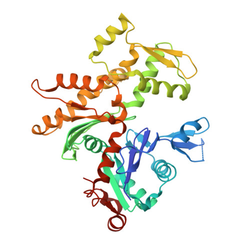
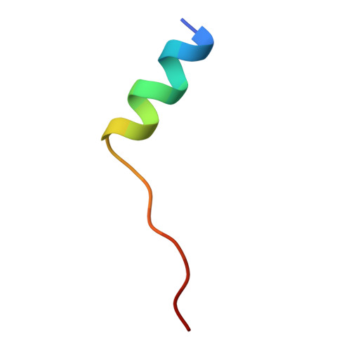
Structures of actin-bound Wiskott-Aldrich syndrome protein homology 2 (WH2) domains of Spire and the implication for filament nucleation.

Ducka, A.M.Joel, P.Popowicz, G.M.Trybus, K.M.Schleicher, M.Noegel, A.A.Huber, R.Holak, T.A.Sitar, T.

(2010) Proc Natl Acad Sci U S A 107: 11757-11762

  • DOI: https://doi.org/10.1073/pnas.1005347107
  • Primary Citation of Related Structures:  
    3MMV, 3MN5, 3MN6, 3MN7, 3MN9

  • PubMed Abstract: 

    Three classes of proteins are known to nucleate new filaments: the Arp2/3 complex, formins, and the third group of proteins that contain ca. 25 amino acid long actin-binding Wiskott-Aldrich syndrome protein homology 2 domains, called the WH2 repeats. Crystal structures of the complexes between the actin-binding WH2 repeats of the Spire protein and actin were determined for the Spire single WH2 domain D, the double (SpirCD), triple (SpirBCD), quadruple (SpirABCD) domains, and an artificial Spire WH2 construct comprising three identical D repeats (SpirDDD). SpirCD represents the minimal functional core of Spire that can nucleate actin filaments. Packing in the crystals of the actin complexes with SpirCD, SpirBCD, SpirABCD, and SpirDDD shows the presence of two types of assemblies, "side-to-side" and "straight-longitudinal," which can serve as actin filament nuclei. The principal feature of these structures is their loose, open conformations, in which the sides of actins that normally constitute the inner interface core of a filament are flipped inside out. These Spire structures are distant from those seen in the filamentous nuclei of Arp2/3, formins, and in the F-actin filament.


  • Organizational Affiliation

    Max-Planck-Institut für Biochemie, 82152 Martinsried, Germany.


Macromolecules
Find similar proteins by:  (by identity cutoff)  |  3D Structure
Entity ID: 1
MoleculeChains Sequence LengthOrganismDetailsImage
Actin-5C374Drosophila melanogasterMutation(s): 2 
Gene Names: Act5CCG4027
UniProt
Find proteins for P10987 (Drosophila melanogaster)
Explore P10987 
Go to UniProtKB:  P10987
Entity Groups  
Sequence Clusters30% Identity50% Identity70% Identity90% Identity95% Identity100% Identity
UniProt GroupP10987
Sequence Annotations
Expand
  • Reference Sequence

Find similar proteins by:  Sequence   |   3D Structure  

Entity ID: 2
MoleculeChains Sequence LengthOrganismDetailsImage
Protein spireB [auth X]19Drosophila melanogasterMutation(s): 0 
Gene Names: spirp150-SpirCG10076
UniProt
Find proteins for Q9U1K1 (Drosophila melanogaster)
Explore Q9U1K1 
Go to UniProtKB:  Q9U1K1
Entity Groups  
UniProt GroupQ9U1K1
Sequence Annotations
Expand
  • Reference Sequence
Small Molecules
Ligands 2 Unique
IDChains Name / Formula / InChI Key2D Diagram3D Interactions
ATP
Query on ATP

Download Ideal Coordinates CCD File 
D [auth A]ADENOSINE-5'-TRIPHOSPHATE
C10 H16 N5 O13 P3
ZKHQWZAMYRWXGA-KQYNXXCUSA-N
CA
Query on CA

Download Ideal Coordinates CCD File 
C [auth A]CALCIUM ION
Ca
BHPQYMZQTOCNFJ-UHFFFAOYSA-N
Experimental Data & Validation

Experimental Data

  • Method: X-RAY DIFFRACTION
  • Resolution: 2.80 Å
  • R-Value Free: 0.330 
  • R-Value Work: 0.241 
  • R-Value Observed: 0.245 
  • Space Group: P 65
Unit Cell:
Length ( Å )Angle ( ˚ )
a = 125.51α = 90
b = 125.51β = 90
c = 56.1γ = 120
Software Package:
Software NamePurpose
MAR345data collection
MOLREPphasing
REFMACrefinement
XDSdata reduction
XSCALEdata scaling

Structure Validation

View Full Validation Report


Ligand Structure Quality Assessment 


Entry History 

Deposition Data

Revision History  (Full details and data files)

  • Version 1.0: 2010-06-02
    Type: Initial release
  • Version 1.1: 2011-07-13
    Changes: Version format compliance
  • Version 1.2: 2017-11-08
    Changes: Refinement description
  • Version 1.3: 2021-10-06
    Changes: Database references, Derived calculations
  • Version 1.4: 2023-09-06
    Changes: Data collection, Refinement description