3MMR

Structure of Plasmodium falciparum Arginase in complex with ABH


Experimental Data Snapshot

  • Method: X-RAY DIFFRACTION
  • Resolution: 2.14 Å
  • R-Value Free: 0.186 
  • R-Value Work: 0.155 
  • R-Value Observed: 0.159 

wwPDB Validation   3D Report Full Report


Ligand Structure Quality Assessment 


This is version 1.2 of the entry. See complete history


Literature

Crystal structure of arginase from Plasmodium falciparum and implications for L-arginine depletion in malarial infection .

Dowling, D.P.Ilies, M.Olszewski, K.L.Portugal, S.Mota, M.M.Llinas, M.Christianson, D.W.

(2010) Biochemistry 49: 5600-5608

  • DOI: https://doi.org/10.1021/bi100390z
  • Primary Citation of Related Structures:  
    3MMR

  • PubMed Abstract: 

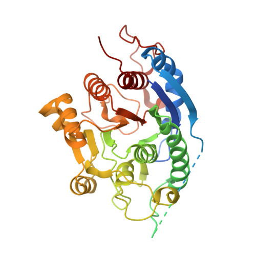
    The 2.15 A resolution crystal structure of arginase from Plasmodium falciparum, the parasite that causes cerebral malaria, is reported in complex with the boronic acid inhibitor 2(S)-amino-6-boronohexanoic acid (ABH) (K(d) = 11 microM). This is the first crystal structure of a parasitic arginase. Various protein constructs were explored to identify an optimally active enzyme form for inhibition and structural studies and to probe the structure and function of two polypeptide insertions unique to malarial arginase: a 74-residue low-complexity region contained in loop L2 and an 11-residue segment contained in loop L8. Structural studies indicate that the low-complexity region is largely disordered and is oriented away from the trimer interface; its deletion does not significantly compromise enzyme activity. The loop L8 insertion is located at the trimer interface and makes several intra- and intermolecular interactions important for enzyme function. In addition, we also demonstrate that arg- Plasmodium berghei sporozoites show significantly decreased liver infectivity in vivo. Therefore, inhibition of malarial arginase may serve as a possible candidate for antimalarial therapy against liver-stage infection, and ABH may serve as a lead for the development of inhibitors.


  • Organizational Affiliation

    Roy and Diana Vagelos Laboratories, Department of Chemistry, University of Pennsylvania, Philadelphia, Pennsylvania 19104-6323, USA.


Macromolecules
Find similar proteins by:  (by identity cutoff)  |  3D Structure
Entity ID: 1
MoleculeChains Sequence LengthOrganismDetailsImage
Arginase413Plasmodium falciparum 3D7Mutation(s): 0 
Gene Names: PFI0320w
EC: 3.5.3.1
UniProt
Find proteins for Q8I384 (Plasmodium falciparum (isolate 3D7))
Explore Q8I384 
Go to UniProtKB:  Q8I384
Entity Groups  
Sequence Clusters30% Identity50% Identity70% Identity90% Identity95% Identity100% Identity
UniProt GroupQ8I384
Sequence Annotations
Expand
  • Reference Sequence
Small Molecules
Binding Affinity Annotations 
IDSourceBinding Affinity
ABH Binding MOAD:  3MMR Kd: 1.10e+4 (nM) from 1 assay(s)
PDBBind:  3MMR Kd: 1.10e+4 (nM) from 1 assay(s)
Experimental Data & Validation

Experimental Data

  • Method: X-RAY DIFFRACTION
  • Resolution: 2.14 Å
  • R-Value Free: 0.186 
  • R-Value Work: 0.155 
  • R-Value Observed: 0.159 
  • Space Group: H 3 2
Unit Cell:
Length ( Å )Angle ( ˚ )
a = 112.594α = 90
b = 112.594β = 90
c = 228.9γ = 120
Software Package:
Software NamePurpose
CBASSdata collection
PHASERphasing
PHENIXrefinement
HKL-2000data reduction
HKL-2000data scaling

Structure Validation

View Full Validation Report


Ligand Structure Quality Assessment 


Entry History 

Deposition Data

Revision History  (Full details and data files)

  • Version 1.0: 2010-06-23
    Type: Initial release
  • Version 1.1: 2011-07-13
    Changes: Version format compliance
  • Version 1.2: 2023-09-06
    Changes: Data collection, Database references, Derived calculations, Refinement description