3MLB

BaNadD in complex with inhibitor 1_02_1


Experimental Data Snapshot

  • Method: X-RAY DIFFRACTION
  • Resolution: 1.80 Å
  • R-Value Free: 0.231 
  • R-Value Work: 0.183 
  • R-Value Observed: 0.186 

wwPDB Validation   3D Report Full Report


Ligand Structure Quality Assessment 


This is version 1.2 of the entry. See complete history


Literature

Complexes of bacterial nicotinate mononucleotide adenylyltransferase with inhibitors: implication for structure-based drug design and improvement.

Huang, N.Kolhatkar, R.Eyobo, Y.Sorci, L.Rodionova, I.Osterman, A.L.Mackerell, A.D.Zhang, H.

(2010) J Med Chem 53: 5229-5239

  • DOI: https://doi.org/10.1021/jm100377f
  • Primary Citation of Related Structures:  
    3MLA, 3MLB, 3MMX

  • PubMed Abstract: 

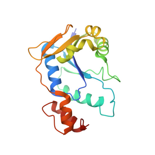
    Bacterial nicotinate mononucleotide adenylyltransferase encoded by the essential gene nadD plays a central role in the synthesis of the redox cofactor NAD(+). The NadD enzyme is conserved in the majority of bacterial species and has been recognized as a novel target for developing new and potentially broad-spectrum antibacterial therapeutics. Here we report the crystal structures of Bacillus anthracis NadD in complex with three NadD inhibitors, including two analogues synthesized in the present study. These structures revealed a common binding site shared by different classes of NadD inhibitors and explored the chemical environment surrounding this site. The structural data obtained here also showed that the subtle changes in ligand structure can lead to significant changes in the binding mode, information that will be useful for future structure-based optimization and design of high affinity inhibitors.


  • Organizational Affiliation

    Department of Biochemistry, University of Texas Southwestern Medical Center, 5323 Harry Hines Boulevard, Dallas, Texas 75390, USA.


Macromolecules
Find similar proteins by:  (by identity cutoff)  |  3D Structure
Entity ID: 1
MoleculeChains Sequence LengthOrganismDetailsImage
nicotinate-nucleotide adenylyltransferase
A, B
191Bacillus anthracisMutation(s): 0 
Gene Names: BAMEG_4595nadD
EC: 2.7.7.18
UniProt
Find proteins for C3L5T6 (Bacillus anthracis (strain CDC 684 / NRRL 3495))
Explore C3L5T6 
Go to UniProtKB:  C3L5T6
Entity Groups  
Sequence Clusters30% Identity50% Identity70% Identity90% Identity95% Identity100% Identity
UniProt GroupC3L5T6
Sequence Annotations
Expand
  • Reference Sequence
Small Molecules
Binding Affinity Annotations 
IDSourceBinding Affinity
LJZ PDBBind:  3MLB IC50: 1.60e+4 (nM) from 1 assay(s)
Binding MOAD:  3MLB IC50: 1.60e+4 (nM) from 1 assay(s)
Experimental Data & Validation

Experimental Data

  • Method: X-RAY DIFFRACTION
  • Resolution: 1.80 Å
  • R-Value Free: 0.231 
  • R-Value Work: 0.183 
  • R-Value Observed: 0.186 
  • Space Group: P 21 21 2
Unit Cell:
Length ( Å )Angle ( ˚ )
a = 88.344α = 90
b = 96.639β = 90
c = 44.132γ = 90
Software Package:
Software NamePurpose
HKL-2000data collection
MOLREPphasing
PHENIXrefinement
HKL-2000data reduction
HKL-2000data scaling

Structure Validation

View Full Validation Report


Ligand Structure Quality Assessment 


Entry History 

Deposition Data

Revision History  (Full details and data files)

  • Version 1.0: 2010-07-28
    Type: Initial release
  • Version 1.1: 2011-07-13
    Changes: Version format compliance
  • Version 1.2: 2023-09-06
    Changes: Data collection, Database references, Derived calculations, Refinement description