3MKQ

Crystal structure of yeast alpha/betaprime-COP subcomplex of the COPI vesicular coat


Experimental Data Snapshot

  • Method: X-RAY DIFFRACTION
  • Resolution: 2.50 Å
  • R-Value Free: 0.272 
  • R-Value Work: 0.221 
  • R-Value Observed: 0.221 

wwPDB Validation   3D Report Full Report


This is version 1.2 of the entry. See complete history


Literature

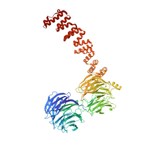
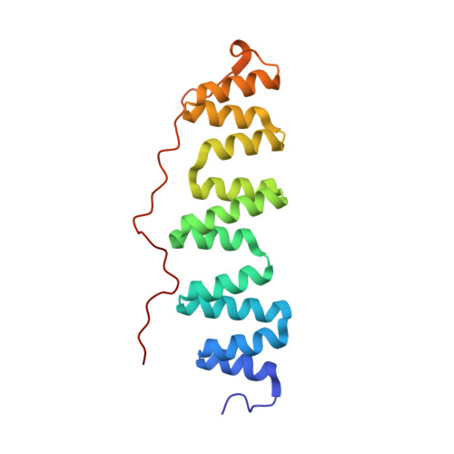
Structure of coatomer cage proteins and the relationship among COPI, COPII, and clathrin vesicle coats.

Lee, C.Goldberg, J.

(2010) Cell 142: 123-132

  • DOI: https://doi.org/10.1016/j.cell.2010.05.030
  • Primary Citation of Related Structures:  
    3MKQ, 3MKR

  • PubMed Abstract: 

    COPI-coated vesicles form at the Golgi apparatus from two cytosolic components, ARF G protein and coatomer, a heptameric complex that can polymerize into a cage to deform the membrane into a bud. Although coatomer shares a common evolutionary origin with COPII and clathrin vesicle coat proteins, the architectural relationship among the three cages is unclear. Strikingly, the alphabeta'-COP core of coatomer crystallizes as a triskelion in which three copies of a beta'-COP beta-propeller domain converge through their axial ends. We infer that the trimer constitutes the vertex of the COPI cage. Our model proposes that the COPI cage is intermediate in design between COPII and clathrin: COPI shares with clathrin an arrangement of three curved alpha-solenoid legs radiating from a common center, and COPI shares with COPII highly similar vertex interactions involving the axial ends of beta-propeller domains.


  • Organizational Affiliation

    Howard Hughes Medical Institute, Memorial Sloan-Kettering Cancer Center, 1275 York Avenue, New York, NY 10065, USA.


Macromolecules
Find similar proteins by:  (by identity cutoff)  |  3D Structure
Entity ID: 1
MoleculeChains Sequence LengthOrganismDetailsImage
Coatomer beta'-subunit
A, C, E
814Saccharomyces cerevisiae YJM789Mutation(s): 1 
Gene Names: SEC27SCY_1929
UniProt
Find proteins for A6ZU46 (Saccharomyces cerevisiae (strain YJM789))
Explore A6ZU46 
Go to UniProtKB:  A6ZU46
Entity Groups  
Sequence Clusters30% Identity50% Identity70% Identity90% Identity95% Identity100% Identity
UniProt GroupA6ZU46
Sequence Annotations
Expand
  • Reference Sequence
Find similar proteins by:  (by identity cutoff)  |  3D Structure
Entity ID: 2
MoleculeChains Sequence LengthOrganismDetailsImage
Coatomer subunit alpha
B, D, F
177Saccharomyces cerevisiaeMutation(s): 2 
Gene Names: RET1COP1SEC33YDL145CD1578
UniProt
Find proteins for P53622 (Saccharomyces cerevisiae (strain ATCC 204508 / S288c))
Explore P53622 
Go to UniProtKB:  P53622
Entity Groups  
Sequence Clusters30% Identity50% Identity70% Identity90% Identity95% Identity100% Identity
UniProt GroupP53622
Sequence Annotations
Expand
  • Reference Sequence
Experimental Data & Validation

Experimental Data

  • Method: X-RAY DIFFRACTION
  • Resolution: 2.50 Å
  • R-Value Free: 0.272 
  • R-Value Work: 0.221 
  • R-Value Observed: 0.221 
  • Space Group: P 32 2 1
Unit Cell:
Length ( Å )Angle ( ˚ )
a = 152.638α = 90
b = 152.638β = 90
c = 294.535γ = 120
Software Package:
Software NamePurpose
HKL-2000data collection
SOLVEphasing
CNSrefinement
HKL-2000data reduction
HKL-2000data scaling

Structure Validation

View Full Validation Report


Entry History 

Deposition Data

Revision History  (Full details and data files)

  • Version 1.0: 2010-07-14
    Type: Initial release
  • Version 1.1: 2011-07-13
    Changes: Source and taxonomy, Version format compliance
  • Version 1.2: 2024-02-21
    Changes: Data collection, Database references