3MKA

Crystal Structure of Mycobacterium Tuberculosis Proteasome with propetide and an T1A mutation at beta-subunit


Experimental Data Snapshot

  • Method: X-RAY DIFFRACTION
  • Resolution: 2.51 Å
  • R-Value Free: 0.247 
  • R-Value Work: 0.217 
  • R-Value Observed: 0.217 

wwPDB Validation   3D Report Full Report


This is version 1.3 of the entry. See complete history


Literature

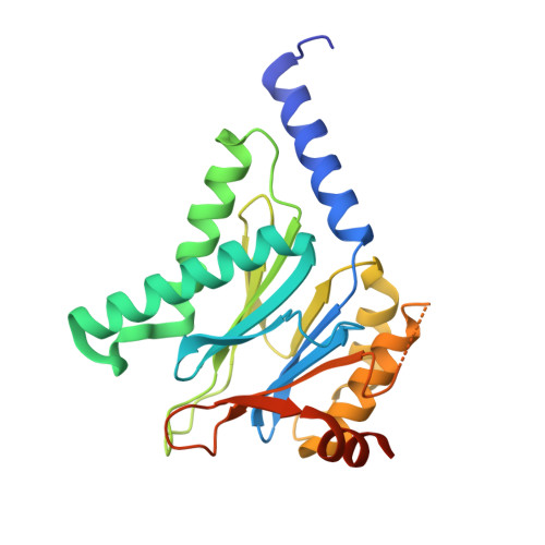
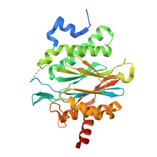
Structural basis for the assembly and gate closure mechanisms of the Mycobacterium tuberculosis 20S proteasome.

Li, D.Li, H.Wang, T.Pan, H.Lin, G.Li, H.

(2010) EMBO J 29: 2037-2047

  • DOI: https://doi.org/10.1038/emboj.2010.95
  • Primary Citation of Related Structures:  
    3MFE, 3MI0, 3MKA

  • PubMed Abstract: 

    Mycobacterium tuberculosis (Mtb) possesses a proteasome system analogous to the eukaryotic ubiquitin-proteasome pathway. Mtb requires the proteasome to resist killing by the host immune system. The detailed assembly process and the gating mechanism of Mtb proteasome have remained unknown. Using cryo-electron microscopy and X-ray crystallography, we have obtained structures of three Mtb proteasome assembly intermediates, showing conformational changes during assembly, and explaining why the beta-subunit propeptide inhibits rather than promotes assembly. Although the eukaryotic proteasome core particles close their protein substrate entrance gates with different amino terminal peptides of the seven alpha-subunits, it has been unknown how a prokaryotic proteasome might close the gate at the symmetry axis with seven identical peptides. We found in the new Mtb proteasome crystal structure that the gate is tightly sealed by the seven identical peptides taking on three distinct conformations. Our work provides the structural bases for assembly and gating mechanisms of the Mtb proteasome.


  • Organizational Affiliation

    Department of Biology, Brookhaven National Laboratory, Upton, NY, USA.


Macromolecules
Find similar proteins by:  (by identity cutoff)  |  3D Structure
Entity ID: 1
MoleculeChains Sequence LengthOrganismDetailsImage
Proteasome subunit alpha
A,
B,
BA [auth 1],
D,
F,
I,
K,
M,
O,
Q,
S,
U,
W,
Y
248Mycobacterium tuberculosisMutation(s): 0 
Gene Names: MT2169prcARv2109c
EC: 3.4.25.1
UniProt
Find proteins for P9WHU1 (Mycobacterium tuberculosis (strain ATCC 25618 / H37Rv))
Explore P9WHU1 
Go to UniProtKB:  P9WHU1
Entity Groups  
Sequence Clusters30% Identity50% Identity70% Identity90% Identity95% Identity100% Identity
UniProt GroupP9WHU1
Sequence Annotations
Expand
  • Reference Sequence
Find similar proteins by:  (by identity cutoff)  |  3D Structure
Entity ID: 2
MoleculeChains Sequence LengthOrganismDetailsImage
Proteasome subunit beta
AA [auth 2],
C,
E,
G,
H,
J,
L,
N,
P,
R,
T,
V,
X,
Z
291Mycobacterium tuberculosisMutation(s): 1 
Gene Names: MT2170prcBRv2110c
EC: 3.4.25.1
UniProt
Find proteins for P9WHT9 (Mycobacterium tuberculosis (strain ATCC 25618 / H37Rv))
Explore P9WHT9 
Go to UniProtKB:  P9WHT9
Entity Groups  
Sequence Clusters30% Identity50% Identity70% Identity90% Identity95% Identity100% Identity
UniProt GroupP9WHT9
Sequence Annotations
Expand
  • Reference Sequence
Experimental Data & Validation

Experimental Data

  • Method: X-RAY DIFFRACTION
  • Resolution: 2.51 Å
  • R-Value Free: 0.247 
  • R-Value Work: 0.217 
  • R-Value Observed: 0.217 
  • Space Group: P 1 21 1
Unit Cell:
Length ( Å )Angle ( ˚ )
a = 173.927α = 90
b = 115.42β = 112.89
c = 199.576γ = 90
Software Package:
Software NamePurpose
CBASSdata collection
CNSrefinement
HKL-2000data reduction
HKL-2000data scaling
CNSphasing

Structure Validation

View Full Validation Report


Entry History 

Deposition Data

  • Released Date: 2010-05-26 
  • Deposition Author(s): Li, D., Li, H.

Revision History  (Full details and data files)

  • Version 1.0: 2010-05-26
    Type: Initial release
  • Version 1.1: 2011-07-13
    Changes: Version format compliance
  • Version 1.2: 2021-10-06
    Changes: Database references
  • Version 1.3: 2023-09-06
    Changes: Data collection, Refinement description