


Crystal Structure of Methyl Transferase from Methanosarcina mazei
Syed Ibrahim, B., Burley, S.K., Swaminathan, S.To be published.
Experimental Data Snapshot
wwPDB Validation 3D Report Full Report

Entity ID: 1 | |||||
|---|---|---|---|---|---|
| Molecule | Chains | Sequence Length | Organism | Details | Image |
| Methyltransferase | 276 | Methanosarcina mazei | Mutation(s): 0 Gene Names: MM_1949 EC: 2.1.1 | 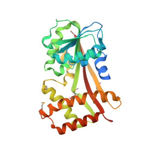 | |
UniProt | |||||
Find proteins for Q8PVL4 (Methanosarcina mazei (strain ATCC BAA-159 / DSM 3647 / Goe1 / Go1 / JCM 11833 / OCM 88)) Explore Q8PVL4 Go to UniProtKB: Q8PVL4 | |||||
Entity Groups | |||||
| Sequence Clusters | 30% Identity50% Identity70% Identity90% Identity95% Identity100% Identity | ||||
| UniProt Group | Q8PVL4 | ||||
Sequence AnnotationsExpand | |||||
| |||||
| Modified Residues 1 Unique | |||||
|---|---|---|---|---|---|
| ID | Chains | Type | Formula | 2D Diagram | Parent |
| MSE Query on MSE | A, B | L-PEPTIDE LINKING | C5 H11 N O2 Se |  | MET |
| Length ( Å ) | Angle ( ˚ ) |
|---|---|
| a = 46.805 | α = 90 |
| b = 56.019 | β = 90 |
| c = 234.607 | γ = 90 |
| Software Name | Purpose |
|---|---|
| CBASS | data collection |
| SHELX | model building |
| SHARP | phasing |
| CNS | refinement |
| HKL-2000 | data reduction |
| SCALEPACK | data scaling |
| SHELX | phasing |
RCSB PDB (citation) is hosted by
RCSB PDB is a member of the

