3MFI

DNA Polymerase Eta in Complex With a cis-syn Thymidine Dimer


Experimental Data Snapshot

  • Method: X-RAY DIFFRACTION
  • Resolution: 1.76 Å
  • R-Value Free: 0.187 
  • R-Value Work: 0.164 
  • R-Value Observed: 0.165 

wwPDB Validation   3D Report Full Report


Ligand Structure Quality Assessment 


This is version 1.4 of the entry. See complete history


Literature

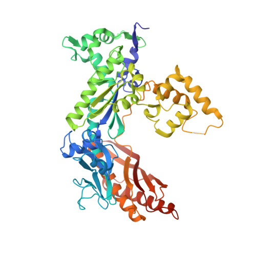
Structural basis for the suppression of skin cancers by DNA polymerase eta.

Silverstein, T.D.Johnson, R.E.Jain, R.Prakash, L.Prakash, S.Aggarwal, A.K.

(2010) Nature 465: 1039-1043

  • DOI: https://doi.org/10.1038/nature09104
  • Primary Citation of Related Structures:  
    3MFH, 3MFI

  • PubMed Abstract: 

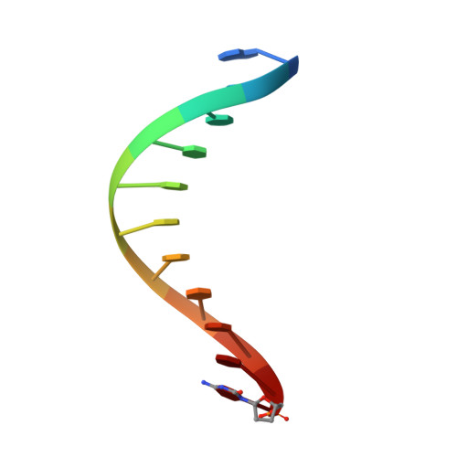
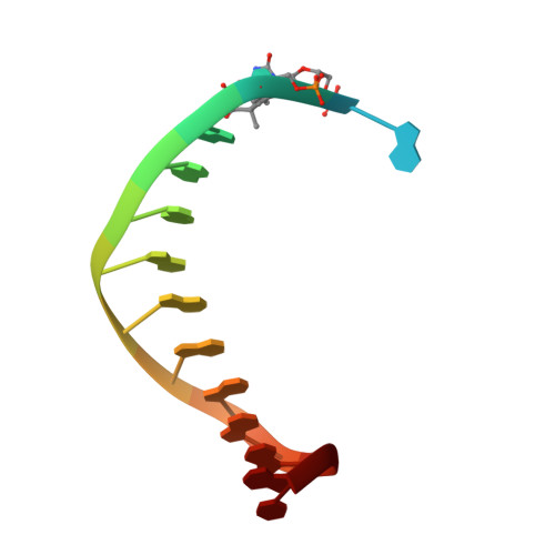
    DNA polymerase eta (Poleta) is unique among eukaryotic polymerases in its proficient ability for error-free replication through ultraviolet-induced cyclobutane pyrimidine dimers, and inactivation of Poleta (also known as POLH) in humans causes the variant form of xeroderma pigmentosum (XPV). We present the crystal structures of Saccharomyces cerevisiae Poleta (also known as RAD30) in ternary complex with a cis-syn thymine-thymine (T-T) dimer and with undamaged DNA. The structures reveal that the ability of Poleta to replicate efficiently through the ultraviolet-induced lesion derives from a simple and yet elegant mechanism, wherein the two Ts of the T-T dimer are accommodated in an active site cleft that is much more open than in other polymerases. We also show by structural, biochemical and genetic analysis that the two Ts are maintained in a stable configuration in the active site via interactions with Gln 55, Arg 73 and Met 74. Together, these features define the basis for Poleta's action on ultraviolet-damaged DNA that is crucial in suppressing the mutagenic and carcinogenic consequences of sun exposure, thereby reducing the incidence of skin cancers in humans.


  • Organizational Affiliation

    Department of Structural and Chemical Biology, Mount Sinai School of Medicine, Box 1677, 1425 Madison Avenue, New York, New York 10029, USA.


Macromolecules

Find similar proteins by:  (by identity cutoff)  |  3D Structure
Entity ID: 1
MoleculeChains Sequence LengthOrganismDetailsImage
DNA polymerase eta520Saccharomyces cerevisiaeMutation(s): 2 
Gene Names: DBH1RAD30YDR419W
EC: 2.7.7.7
UniProt
Find proteins for Q04049 (Saccharomyces cerevisiae (strain ATCC 204508 / S288c))
Explore Q04049 
Go to UniProtKB:  Q04049
Entity Groups  
Sequence Clusters30% Identity50% Identity70% Identity90% Identity95% Identity100% Identity
UniProt GroupQ04049
Sequence Annotations
Expand
  • Reference Sequence

Find similar nucleic acids by:  Sequence   |   3D Structure  

Entity ID: 2
MoleculeChains LengthOrganismImage
5'-D(*GP*TP*CP*CP*TP*CP*CP*CP*CP*TP*(DOC))-3'B [auth P]11N/A
Sequence Annotations
Expand
  • Reference Sequence

Find similar nucleic acids by:  Sequence   |   3D Structure  

Entity ID: 3
MoleculeChains LengthOrganismImage
5'-D(*TP*AP*AP*(TTD)P*GP*AP*GP*GP*GP*GP*AP*GP*GP*AP*C)-3'C [auth T]15N/A
Sequence Annotations
Expand
  • Reference Sequence
Experimental Data & Validation

Experimental Data

  • Method: X-RAY DIFFRACTION
  • Resolution: 1.76 Å
  • R-Value Free: 0.187 
  • R-Value Work: 0.164 
  • R-Value Observed: 0.165 
  • Space Group: C 2 2 21
Unit Cell:
Length ( Å )Angle ( ˚ )
a = 88.087α = 90
b = 226.962β = 90
c = 85.919γ = 90
Software Package:
Software NamePurpose
DENZOdata reduction
SCALEPACKdata scaling
REFMACrefinement
PDB_EXTRACTdata extraction
ADSCdata collection
PHASERphasing

Structure Validation

View Full Validation Report


Ligand Structure Quality Assessment 


Entry History 

Deposition Data

Revision History  (Full details and data files)

  • Version 1.0: 2010-06-23
    Type: Initial release
  • Version 1.1: 2011-07-13
    Changes: Version format compliance
  • Version 1.2: 2017-11-08
    Changes: Refinement description
  • Version 1.3: 2021-10-06
    Changes: Database references, Derived calculations, Structure summary
  • Version 1.4: 2023-09-06
    Changes: Data collection, Refinement description