3MDC

DNA polymerase lambda in complex with dFdCTP


Experimental Data Snapshot

  • Method: X-RAY DIFFRACTION
  • Resolution: 2.00 Å
  • R-Value Free: 0.266 
  • R-Value Work: 0.216 
  • R-Value Observed: 0.219 

wwPDB Validation   3D Report Full Report


Ligand Structure Quality Assessment 


This is version 1.3 of the entry. See complete history


Literature

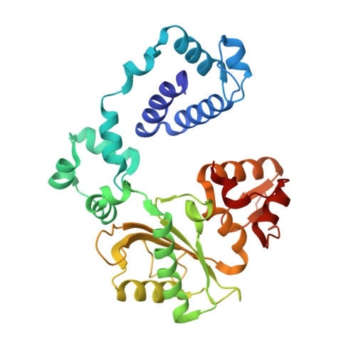
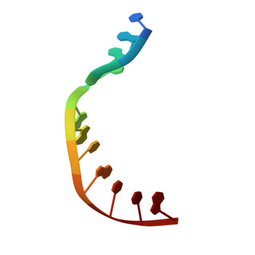
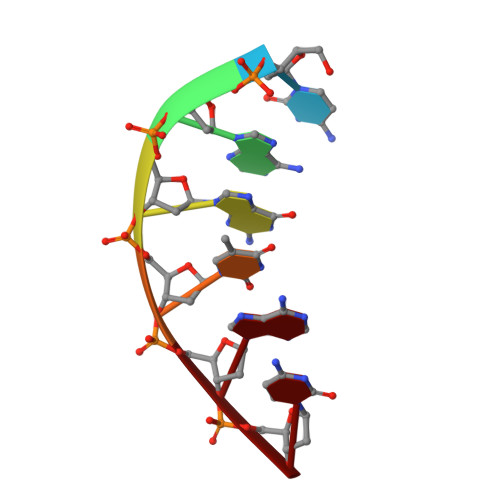
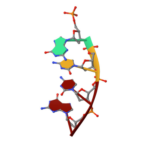
Interaction between DNA Polymerase lambda and anticancer nucleoside analogs.

Garcia-Diaz, M.Murray, M.S.Kunkel, T.A.Chou, K.M.

(2010) J Biol Chem 285: 16874-16879

  • DOI: https://doi.org/10.1074/jbc.M109.094391
  • Primary Citation of Related Structures:  
    3MDA, 3MDC

  • PubMed Abstract: 

    The anticancer activity of cytarabine (AraC) and gemcitabine (dFdC) is thought to result from chain termination after incorporation into DNA. To investigate their incorporation into DNA at atomic level resolution, we present crystal structures of human DNA polymerase lambda (Pol lambda) bound to gapped DNA and containing either AraC or dFdC paired opposite template dG. These structures reveal that AraC and dFdC can bind within the nascent base pair binding pocket of Pol lambda. Although the conformation of the ribose of AraCTP is similar to that of normal dCTP, the conformation of dFdCTP is significantly different. Consistent with these structures, Pol lambda efficiently incorporates AraCTP but not dFdCTP. The data are consistent with the possibility that Pol lambda could modulate the cytotoxic effect of AraC.


  • Organizational Affiliation

    Laboratory of Structural Biology and Laboratory of Molecular Genetics, Department of Health and Human Services, NIEHS, National Institutes of Health, Research Triangle Park, North Carolina 27709, USA.


Macromolecules

Find similar proteins by:  (by identity cutoff)  |  3D Structure
Entity ID: 1
MoleculeChains Sequence LengthOrganismDetailsImage
DNA polymerase lambda325Homo sapiensMutation(s): 0 
Gene Names: POLL
EC: 2.7.7.7 (PDB Primary Data), 4.2.99 (PDB Primary Data)
UniProt & NIH Common Fund Data Resources
Find proteins for Q9UGP5 (Homo sapiens)
Explore Q9UGP5 
Go to UniProtKB:  Q9UGP5
PHAROS:  Q9UGP5
GTEx:  ENSG00000166169 
Entity Groups  
Sequence Clusters30% Identity50% Identity70% Identity90% Identity95% Identity100% Identity
UniProt GroupQ9UGP5
Sequence Annotations
Expand
  • Reference Sequence

Find similar nucleic acids by:  Sequence   |   3D Structure  

Entity ID: 2
MoleculeChains LengthOrganismImage
DNA (5'-D(*CP*GP*GP*CP*GP*GP*TP*AP*CP*TP*G)-3')B [auth T]11N/A
Sequence Annotations
Expand
  • Reference Sequence

Find similar nucleic acids by:  Sequence   |   3D Structure  

Entity ID: 3
MoleculeChains LengthOrganismImage
DNA (5'-D(*CP*AP*GP*TP*AP*C)-3')C [auth P]6N/A
Sequence Annotations
Expand
  • Reference Sequence

Find similar nucleic acids by:  Sequence   |   3D Structure  

Entity ID: 4
MoleculeChains LengthOrganismImage
DNA (5'-D(P*GP*CP*CP*G)-3')4N/A
Sequence Annotations
Expand
  • Reference Sequence
Experimental Data & Validation

Experimental Data

  • Method: X-RAY DIFFRACTION
  • Resolution: 2.00 Å
  • R-Value Free: 0.266 
  • R-Value Work: 0.216 
  • R-Value Observed: 0.219 
  • Space Group: P 21 21 21
Unit Cell:
Length ( Å )Angle ( ˚ )
a = 55.206α = 90
b = 59.773β = 90
c = 142.008γ = 90
Software Package:
Software NamePurpose
DENZOdata reduction
SCALEPACKdata scaling
MOLREPphasing
PHENIXrefinement
PDB_EXTRACTdata extraction

Structure Validation

View Full Validation Report


Ligand Structure Quality Assessment 


Entry History 

Deposition Data

Revision History  (Full details and data files)

  • Version 1.0: 2010-04-28
    Type: Initial release
  • Version 1.1: 2011-07-13
    Changes: Version format compliance
  • Version 1.2: 2017-11-08
    Changes: Refinement description
  • Version 1.3: 2024-02-21
    Changes: Data collection, Database references, Derived calculations, Structure summary