3M6M

Crystal structure of RpfF complexed with REC domain of RpfC


Experimental Data Snapshot

  • Method: X-RAY DIFFRACTION
  • Resolution: 2.50 Å
  • R-Value Free: 0.271 
  • R-Value Work: 0.250 
  • R-Value Observed: 0.251 

wwPDB Validation   3D Report Full Report


This is version 1.3 of the entry. See complete history


Literature

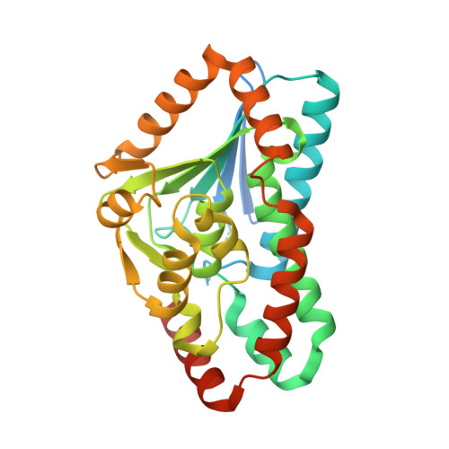
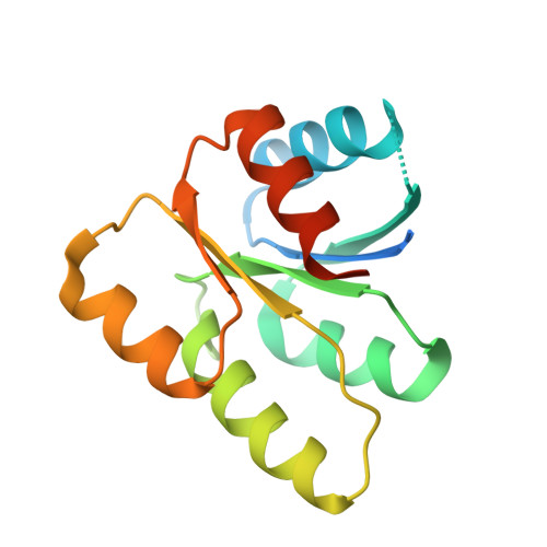
Structural Basis of the Sensor-Synthase Interaction in Autoinduction of the Quorum Sensing Signal DSF Biosynthesis

Cheng, Z.He, Y.W.Lim, S.C.Qamra, R.Walsh, M.A.Zhang, L.H.Song, H.

(2010) Structure 18: 1199-1209

  • DOI: https://doi.org/10.1016/j.str.2010.06.011
  • Primary Citation of Related Structures:  
    3M6M, 3M6N

  • PubMed Abstract: 

    The diffusible signal factor (DSF)-dependent quorum sensing (QS) system adopts a novel protein-protein interaction mechanism to autoregulate the production of signal DSF. Here, we present the crystal structures of DSF synthase RpfF and its complex with the REC domain of sensor protein RpfC. RpfF is structurally similarity to the members of the crotonase superfamily and contains an N-terminal α/β spiral core domain and a C-terminal α-helical region. Further structural and mutational analysis identified two catalytic glutamate residues, which is the conserved feature of the enoyl-CoA hydratases/dehydratases. A putative substrate-binding pocket was unveiled and the key roles of the residues implicated in substrate binding were verified by mutational analysis. The binding of the REC domain may lock RpfF in an inactive conformation by blocking the entrance of substrate binding pocket, thereby negatively regulating DSF production. These findings provide a structural model for the RpfC-RpfF interaction-mediated QS autoinduction mechanism.


  • Organizational Affiliation

    Institute of Molecular and Cell Biology, 61 Biopolis Drive, Proteos, Singapore.


Macromolecules
Find similar proteins by:  (by identity cutoff)  |  3D Structure
Entity ID: 1
MoleculeChains Sequence LengthOrganismDetailsImage
RpfF protein
A, B, C
305Xanthomonas campestris pv. campestrisMutation(s): 0 
Gene Names: rpfFXCC1857
UniProt
Find proteins for Q7CLS3 (Xanthomonas campestris pv. campestris (strain ATCC 33913 / DSM 3586 / NCPPB 528 / LMG 568 / P 25))
Explore Q7CLS3 
Go to UniProtKB:  Q7CLS3
Entity Groups  
Sequence Clusters30% Identity50% Identity70% Identity90% Identity95% Identity100% Identity
UniProt GroupQ7CLS3
Sequence Annotations
Expand
  • Reference Sequence
Find similar proteins by:  (by identity cutoff)  |  3D Structure
Entity ID: 2
MoleculeChains Sequence LengthOrganismDetailsImage
Sensory/regulatory protein rpfC
D, E, F
143Xanthomonas campestris pv. campestrisMutation(s): 0 
Gene Names: rpfCXCC1856
EC: 2.7.13.3
UniProt
Find proteins for P0C0F6 (Xanthomonas campestris pv. campestris (strain ATCC 33913 / DSM 3586 / NCPPB 528 / LMG 568 / P 25))
Explore P0C0F6 
Go to UniProtKB:  P0C0F6
Entity Groups  
Sequence Clusters30% Identity50% Identity70% Identity90% Identity95% Identity100% Identity
UniProt GroupP0C0F6
Sequence Annotations
Expand
  • Reference Sequence
Small Molecules
Ligands 3 Unique
IDChains Name / Formula / InChI Key2D Diagram3D Interactions
IOD
Query on IOD

Download Ideal Coordinates CCD File 
G [auth A]
I [auth A]
J [auth B]
K [auth B]
M [auth C]
G [auth A],
I [auth A],
J [auth B],
K [auth B],
M [auth C],
N [auth C],
P [auth C],
Q [auth C],
R [auth D],
S [auth D],
U [auth E],
V [auth E],
X [auth F],
Y [auth F]
IODIDE ION
I
XMBWDFGMSWQBCA-UHFFFAOYSA-M
EDO
Query on EDO

Download Ideal Coordinates CCD File 
H [auth A],
L [auth B],
O [auth C]
1,2-ETHANEDIOL
C2 H6 O2
LYCAIKOWRPUZTN-UHFFFAOYSA-N
MG
Query on MG

Download Ideal Coordinates CCD File 
T [auth D],
W [auth E],
Z [auth F]
MAGNESIUM ION
Mg
JLVVSXFLKOJNIY-UHFFFAOYSA-N
Experimental Data & Validation

Experimental Data

  • Method: X-RAY DIFFRACTION
  • Resolution: 2.50 Å
  • R-Value Free: 0.271 
  • R-Value Work: 0.250 
  • R-Value Observed: 0.251 
  • Space Group: P 65
Unit Cell:
Length ( Å )Angle ( ˚ )
a = 130.913α = 90
b = 130.913β = 90
c = 156.471γ = 120
Software Package:
Software NamePurpose
PHASERphasing
REFMACrefinement
PDB_EXTRACTdata extraction
DNAdata collection
MOSFLMdata reduction
SCALAdata scaling

Structure Validation

View Full Validation Report


Entry History 

Deposition Data

Revision History  (Full details and data files)

  • Version 1.0: 2010-09-22
    Type: Initial release
  • Version 1.1: 2011-07-13
    Changes: Version format compliance
  • Version 1.2: 2013-10-16
    Changes: Derived calculations
  • Version 1.3: 2024-03-20
    Changes: Data collection, Database references, Derived calculations, Refinement description