3M6B

Crystal Structure of the Ertapenem Pre-isomerized Covalent Adduct with TB B-lactamase


Experimental Data Snapshot

  • Method: X-RAY DIFFRACTION
  • Resolution: 1.30 Å
  • R-Value Free: 0.176 
  • R-Value Work: 0.147 
  • R-Value Observed: 0.149 

wwPDB Validation   3D Report Full Report


Ligand Structure Quality Assessment 


This is version 1.4 of the entry. See complete history


Literature

Biochemical and structural characterization of Mycobacterium tuberculosis beta-lactamase with the carbapenems ertapenem and doripenem.

Tremblay, L.W.Fan, F.Blanchard, J.S.

(2010) Biochemistry 49: 3766-3773

  • DOI: https://doi.org/10.1021/bi100232q
  • Primary Citation of Related Structures:  
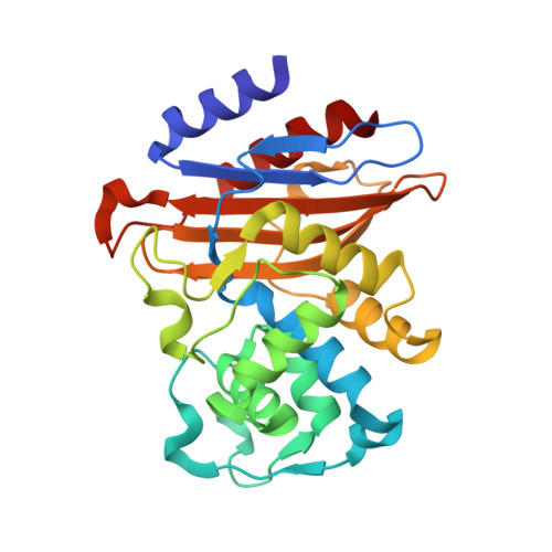
    3IQA, 3M6B, 3M6H

  • PubMed Abstract: 

    Despite the enormous success of beta-lactams as broad-spectrum antibacterials, they have never been widely used for the treatment of tuberculosis (TB) due to intrinsic resistance that is caused by the presence of a chromosomally encoded gene (blaC) in Mycobacterium tuberculosis. Our previous studies of TB BlaC revealed that this enzyme is an extremely broad-spectrum beta-lactamase hydrolyzing all beta-lactam classes. Carbapenems are slow substrates that acylate the enzyme but are only slowly deacylated and can therefore act also as potent inhibitors of BlaC. We conducted the in vitro characterization of doripenem and ertapenem with BlaC. A steady-state kinetic burst was observed with both compounds with magnitudes proportional to the concentration of BlaC used. The results provide apparent K(m) and k(cat) values of 0.18 microM and 0.016 min(-1) for doripenem and 0.18 microM and 0.017 min(-1) for ertapenem, respectively. FTICR mass spectrometry demonstrated that the doripenem and ertapenem acyl-enzyme complexes remain stable over a time period of 90 min. The BlaC-doripenem covalent complex obtained after a 90 min soak was determined to 2.2 A, while the BlaC-ertapenem complex obtained after a 90 min soak was determined to 2.0 A. The 1.3 A diffraction data from a 10 min ertapenem-soaked crystal revealed an isomerization occurring in the BlaC-ertapenem adduct in which the original Delta(2)-pyrroline ring was tautomerized to generate the Delta(1)-pyrroline ring. The isomerization leads to the flipping of the carbapenem hydroxyethyl group to hydrogen bond to carboxyl O2 of Glu166. The hydroxyethyl flip results in both the decreased basicity of Glu166 and a significant increase in the distance between carboxyl O2 of Glu166 and the catalytic water molecule, slowing hydrolysis.


  • Organizational Affiliation

    Department of Biochemistry, Albert Einstein College of Medicine, 1300 Morris Park Avenue, Bronx, New York 10461, USA.


Macromolecules
Find similar proteins by:  (by identity cutoff)  |  3D Structure
Entity ID: 1
MoleculeChains Sequence LengthOrganismDetailsImage
Beta-lactamase265Mycobacterium tuberculosisMutation(s): 0 
Gene Names: blaAblaCRv2068cMT2128MTCY49.07c
EC: 3.5.2.6
UniProt
Find proteins for P9WKD3 (Mycobacterium tuberculosis (strain ATCC 25618 / H37Rv))
Explore P9WKD3 
Go to UniProtKB:  P9WKD3
Entity Groups  
Sequence Clusters30% Identity50% Identity70% Identity90% Identity95% Identity100% Identity
UniProt GroupP9WKD3
Sequence Annotations
Expand
  • Reference Sequence
Small Molecules
Ligands 2 Unique
IDChains Name / Formula / InChI Key2D Diagram3D Interactions
1RG
Query on 1RG

Download Ideal Coordinates CCD File 
D [auth A],
E [auth A]
(4R,5S)-3-({(3S,5S)-5-[(3-carboxyphenyl)carbamoyl]pyrrolidin-3-yl}sulfanyl)-5-[(1S,2R)-1-formyl-2-hydroxypropyl]-4-methyl-4,5-dihydro-1H-pyrrole-2-carboxylic acid
C22 H27 N3 O7 S
PGRRQYXTRXQDDJ-SKHPLXCOSA-N
PO4
Query on PO4

Download Ideal Coordinates CCD File 
B [auth A],
C [auth A]
PHOSPHATE ION
O4 P
NBIIXXVUZAFLBC-UHFFFAOYSA-K
Experimental Data & Validation

Experimental Data

  • Method: X-RAY DIFFRACTION
  • Resolution: 1.30 Å
  • R-Value Free: 0.176 
  • R-Value Work: 0.147 
  • R-Value Observed: 0.149 
  • Space Group: P 21 21 21
Unit Cell:
Length ( Å )Angle ( ˚ )
a = 49.657α = 90
b = 67.922β = 90
c = 75.546γ = 90
Software Package:
Software NamePurpose
REFMACrefinement
PDB_EXTRACTdata extraction
CBASSdata collection
DENZOdata reduction
SCALEPACKdata scaling

Structure Validation

View Full Validation Report


Ligand Structure Quality Assessment 


Entry History 

Deposition Data

Revision History  (Full details and data files)

  • Version 1.0: 2010-04-14
    Type: Initial release
  • Version 1.1: 2011-07-13
    Changes: Version format compliance
  • Version 1.2: 2012-05-16
    Changes: Non-polymer description, Structure summary
  • Version 1.3: 2017-11-08
    Changes: Refinement description
  • Version 1.4: 2023-09-06
    Changes: Data collection, Database references, Derived calculations, Refinement description