3M4V

Crystal structure of the A330P mutant of cytochrome P450 BM3


Experimental Data Snapshot

  • Method: X-RAY DIFFRACTION
  • Resolution: 1.90 Å
  • R-Value Free: 0.232 
  • R-Value Work: 0.187 
  • R-Value Observed: 0.189 

wwPDB Validation   3D Report Full Report


Ligand Structure Quality Assessment 


This is version 1.3 of the entry. See complete history


Literature

Structural basis for the properties of two single-site proline mutants of CYP102A1 (P450BM3)

Whitehouse, C.J.C.Yang, W.Yorke, J.A.Rowlatt, B.C.Strong, A.J.Blanford, C.F.Bell, S.G.Bartlam, M.Wong, L.L.Rao, Z.

(2010) Chembiochem 11: 2549-2556

  • DOI: https://doi.org/10.1002/cbic.201000421
  • Primary Citation of Related Structures:  
    3M4V

  • PubMed Abstract: 

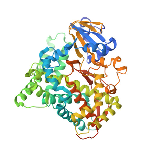
    The crystal structures of the haem domains of Ala330Pro and Ile401Pro, two single-site proline variants of CYP102A1 (P450(BM3)) from Bacillus megaterium, have been solved. In the A330P structure, the active site is constricted by the relocation of the Pro329 side chain into the substrate access channel, providing a basis for the distinctive C-H bond oxidation profiles given by the variant and the enhanced activity with small molecules. I401P, which is exceptionally active towards non-natural substrates, displays a number of structural similarities to substrate-bound forms of the wild-type enzyme, notably an off-axial water ligand, a drop in the proximal loop, and the positioning of two I-helix residues, Gly265 and His266, the reorientation of which prevents the formation of several intrahelical hydrogen bonds. Second-generation I401P variants gave high in vitro oxidation rates with non-natural substrates as varied as fluorene and propane, towards which the wild-type enzyme is essentially inactive. The substrate-free I401P haem domain had a reduction potential slightly more oxidising than the palmitate-bound wild-type haem domain, and a first electron transfer rate that was about 10 % faster. The electronic properties of A330P were, by contrast, similar to those of the substrate-free wild-type enzyme.


  • Organizational Affiliation

    Department of Chemistry, University of Oxford, Inorganic Chemistry Laboratory, South Parks Road, Oxford OX1 3QR, UK.


Macromolecules
Find similar proteins by:  (by identity cutoff)  |  3D Structure
Entity ID: 1
MoleculeChains Sequence LengthOrganismDetailsImage
Bifunctional P-450/NADPH-P450 reductase
A, B
482Priestia megateriumMutation(s): 1 
Gene Names: p450bm-3
EC: 1.14.14.1
UniProt
Find proteins for P14779 (Priestia megaterium (strain ATCC 14581 / DSM 32 / CCUG 1817 / JCM 2506 / NBRC 15308 / NCIMB 9376 / NCTC 10342 / NRRL B-14308 / VKM B-512 / Ford 19))
Explore P14779 
Go to UniProtKB:  P14779
Entity Groups  
Sequence Clusters30% Identity50% Identity70% Identity90% Identity95% Identity100% Identity
UniProt GroupP14779
Sequence Annotations
Expand
  • Reference Sequence
Small Molecules
Ligands 1 Unique
IDChains Name / Formula / InChI Key2D Diagram3D Interactions
HEM
Query on HEM

Download Ideal Coordinates CCD File 
C [auth A],
D [auth B]
PROTOPORPHYRIN IX CONTAINING FE
C34 H32 Fe N4 O4
KABFMIBPWCXCRK-RGGAHWMASA-L
Experimental Data & Validation

Experimental Data

  • Method: X-RAY DIFFRACTION
  • Resolution: 1.90 Å
  • R-Value Free: 0.232 
  • R-Value Work: 0.187 
  • R-Value Observed: 0.189 
  • Space Group: P 1 21 1
Unit Cell:
Length ( Å )Angle ( ˚ )
a = 58.542α = 90
b = 148.281β = 99.93
c = 64.368γ = 90
Software Package:
Software NamePurpose
ADSCdata collection
PHASERphasing
REFMACrefinement
HKL-2000data reduction
HKL-2000data scaling

Structure Validation

View Full Validation Report


Ligand Structure Quality Assessment 


Entry History 

Deposition Data

Revision History  (Full details and data files)

  • Version 1.0: 2011-03-23
    Type: Initial release
  • Version 1.1: 2011-07-13
    Changes: Version format compliance
  • Version 1.2: 2013-12-18
    Changes: Database references
  • Version 1.3: 2023-11-01
    Changes: Data collection, Database references, Derived calculations, Refinement description