3M09

F98Y TMP-resistant Dihydrofolate Reductase from Staphylococcus aureus with inhibitor RAB1


Experimental Data Snapshot

  • Method: X-RAY DIFFRACTION
  • Resolution: 2.01 Å
  • R-Value Free: 0.207 
  • R-Value Work: 0.169 
  • R-Value Observed: 0.173 

wwPDB Validation   3D Report Full Report


Ligand Structure Quality Assessment 


This is version 1.3 of the entry. See complete history


Literature

Inhibition of Antibiotic-Resistant Staphylococcus aureus by the Broad-Spectrum Dihydrofolate Reductase Inhibitor RAB1.

Bourne, C.R.Barrow, E.W.Bunce, R.A.Bourne, P.C.Berlin, K.D.Barrow, W.W.

(2010) Antimicrob Agents Chemother 54: 3825-3833

  • DOI: https://doi.org/10.1128/AAC.00361-10
  • Primary Citation of Related Structures:  
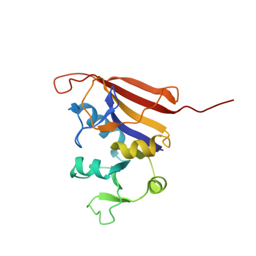
    3M08, 3M09

  • PubMed Abstract: 

    The bacterial burden on human health is quickly outweighing available therapeutics. Our long-term goal is the development of antimicrobials with the potential for broad-spectrum activity. We previously reported phthalazine-based inhibitors of dihydrofolate reductase (DHFR) with potent activity against Bacillus anthracis, a major component of Project BioShield. The most active molecule, named RAB1, performs well in vitro and, in a cocrystal structure, was found deep within the active site of B. anthracis DHFR. We have now examined the activity of RAB1 against a panel of bacteria relevant to human health and found broad-spectrum applicability, particularly with regard to gram-positive organisms. RAB1 was most effective against Staphylococcus aureus, including methicillin- and vancomycin-resistant (MRSA/VRSA) strains. We have determined the cocrystal structure of the wild-type and trimethoprim-resistant (Phe 98 Tyr) DHFR enzyme from S. aureus with RAB1, and we found that rotational freedom of the acryloyl linker region allows the phthalazine moiety to occupy two conformations. This freedom in placement also allows either enantiomer of RAB1 to bind to S. aureus, in contrast to the specificity of B. anthracis for the S-enantiomer. Additionally, one of the conformations of RAB1 defines a unique surface cavity that increases the strength of interaction with S. aureus. These observations provide insights into the binding capacity of S. aureus DHFR and highlight atypical features critical for future exploitation in drug development.


  • Organizational Affiliation

    Department of Veterinary Pathobiology, 250 McElroy Hall, Oklahoma State University, Stillwater, OK 74078, USA. christina.bourne@okstate.edu


Macromolecules
Find similar proteins by:  (by identity cutoff)  |  3D Structure
Entity ID: 1
MoleculeChains Sequence LengthOrganismDetailsImage
Dihydrofolate reductase161Staphylococcus aureusMutation(s): 1 
Gene Names: folA
EC: 1.5.1.3
UniProt
Find proteins for P0A017 (Staphylococcus aureus)
Explore P0A017 
Go to UniProtKB:  P0A017
Entity Groups  
Sequence Clusters30% Identity50% Identity70% Identity90% Identity95% Identity100% Identity
UniProt GroupP0A017
Sequence Annotations
Expand
  • Reference Sequence
Small Molecules
Ligands 3 Unique
IDChains Name / Formula / InChI Key2D Diagram3D Interactions
NAP
Query on NAP

Download Ideal Coordinates CCD File 
C [auth A]NADP NICOTINAMIDE-ADENINE-DINUCLEOTIDE PHOSPHATE
C21 H28 N7 O17 P3
XJLXINKUBYWONI-NNYOXOHSSA-N
RAR
Query on RAR

Download Ideal Coordinates CCD File 
B [auth A]5-(3,4-dimethoxy-5-{(1E)-3-oxo-3-[(1S)-1-propylphthalazin-2(1H)-yl]prop-1-en-1-yl}benzyl)pyrimidine-2,4-diamine
C27 H30 N6 O3
YGDVMSPWZQHNMB-NEQMZLFVSA-N
GOL
Query on GOL

Download Ideal Coordinates CCD File 
D [auth A]GLYCEROL
C3 H8 O3
PEDCQBHIVMGVHV-UHFFFAOYSA-N
Binding Affinity Annotations 
IDSourceBinding Affinity
RAR Binding MOAD:  3M09 Ki: 0.9 (nM) from 1 assay(s)
BindingDB:  3M09 Ki: 0.9 (nM) from 1 assay(s)
IC50: min: 2.9, max: 12.5 (nM) from 2 assay(s)
Experimental Data & Validation

Experimental Data

  • Method: X-RAY DIFFRACTION
  • Resolution: 2.01 Å
  • R-Value Free: 0.207 
  • R-Value Work: 0.169 
  • R-Value Observed: 0.173 
  • Space Group: P 61 2 2
Unit Cell:
Length ( Å )Angle ( ˚ )
a = 79.18α = 90
b = 79.18β = 90
c = 107.88γ = 120
Software Package:
Software NamePurpose
MOSFLMdata reduction
SCALAdata scaling
PHASERphasing
PHENIXrefinement
PDB_EXTRACTdata extraction

Structure Validation

View Full Validation Report


Ligand Structure Quality Assessment 


Entry History 

Deposition Data

Revision History  (Full details and data files)

  • Version 1.0: 2010-07-14
    Type: Initial release
  • Version 1.1: 2011-07-13
    Changes: Version format compliance
  • Version 1.2: 2021-10-13
    Changes: Database references, Derived calculations
  • Version 1.3: 2024-02-21
    Changes: Data collection