3LXU

Crystal Structure of Tripeptidyl Peptidase 2 (TPP II)


Experimental Data Snapshot

  • Method: X-RAY DIFFRACTION
  • Resolution: 3.14 Å
  • R-Value Free: 0.293 
  • R-Value Work: 0.241 

wwPDB Validation   3D Report Full Report


This is version 1.1 of the entry. See complete history


Literature

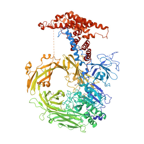
Hybrid molecular structure of the giant protease tripeptidyl peptidase II.

Chuang, C.K.Rockel, B.Seyit, G.Walian, P.J.Schonegge, A.M.Peters, J.Zwart, P.H.Baumeister, W.Jap, B.K.

(2010) Nat Struct Mol Biol 17: 990-996

  • DOI: https://doi.org/10.1038/nsmb.1870
  • Primary Citation of Related Structures:  
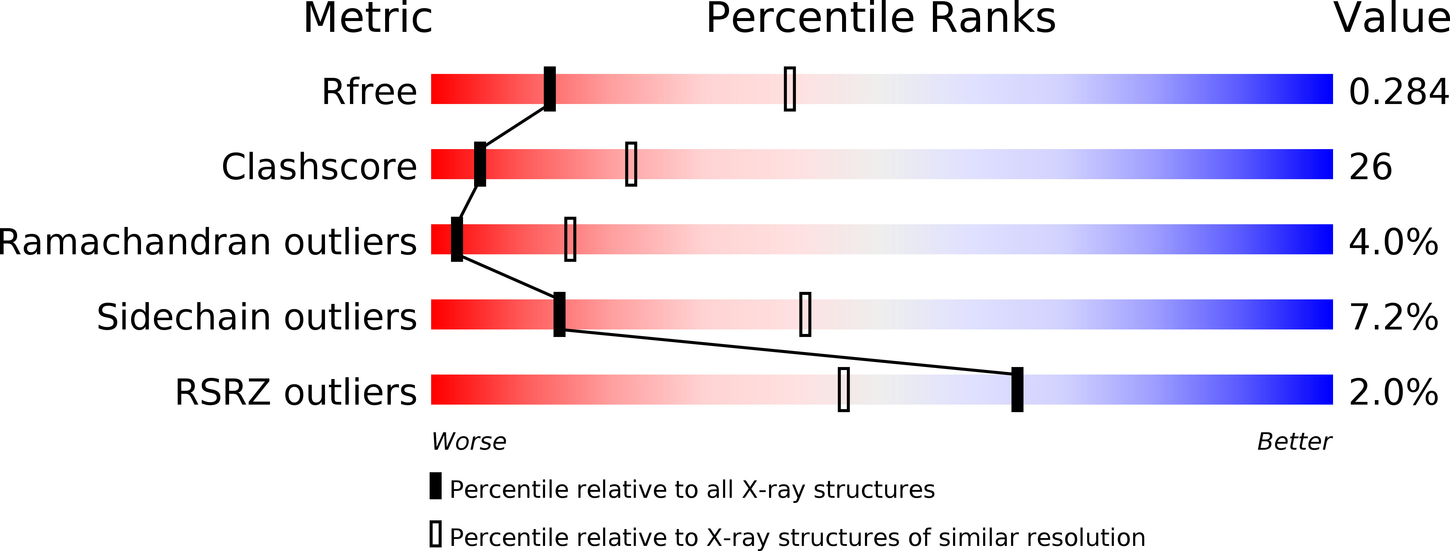
    3LXU

  • PubMed Abstract: 

    Tripeptidyl peptidase II (TPP II) is the largest known eukaryotic protease (6 MDa). It is believed to act downstream of the 26S proteasome, cleaving tripeptides from the N termini of longer peptides, and it is implicated in numerous cellular processes. Here we report the structure of Drosophila TPP II determined by a hybrid approach. We solved the structure of the dimer by X-ray crystallography and docked it into the three-dimensional map of the holocomplex, which we obtained by single-particle cryo-electron microscopy. The resulting structure reveals the compartmentalization of the active sites inside a system of chambers and suggests the existence of a molecular ruler determining the size of the cleavage products. Furthermore, the structure suggests a model for activation of TPP II involving the relocation of a flexible loop and a repositioning of the active-site serine, coupling it to holocomplex assembly and active-site sequestration.


  • Organizational Affiliation

    Life Sciences Division, Lawrence Berkeley National Laboratory, Berkeley, California, USA.


Macromolecules
Find similar proteins by:  (by identity cutoff)  |  3D Structure
Entity ID: 1
MoleculeChains Sequence LengthOrganismDetailsImage
Tripeptidyl-peptidase 2A [auth X]1,354Drosophila melanogasterMutation(s): 0 
Gene Names: CG3991TPP IITppII
EC: 3.4.14.10
UniProt
Find proteins for Q9V6K1 (Drosophila melanogaster)
Explore Q9V6K1 
Go to UniProtKB:  Q9V6K1
Entity Groups  
Sequence Clusters30% Identity50% Identity70% Identity90% Identity95% Identity100% Identity
UniProt GroupQ9V6K1
Sequence Annotations
Expand
  • Reference Sequence
Small Molecules
Modified Residues  1 Unique
IDChains TypeFormula2D DiagramParent
MSE
Query on MSE
A [auth X]L-PEPTIDE LINKINGC5 H11 N O2 SeMET
Experimental Data & Validation

Experimental Data

  • Method: X-RAY DIFFRACTION
  • Resolution: 3.14 Å
  • R-Value Free: 0.293 
  • R-Value Work: 0.241 
  • Space Group: P 41 21 2
Unit Cell:
Length ( Å )Angle ( ˚ )
a = 126.372α = 90
b = 126.372β = 90
c = 213.983γ = 90
Software Package:
Software NamePurpose
ADSCdata collection
PHENIXmodel building
PHENIXrefinement
HKL-2000data reduction
SCALEPACKdata scaling
PHENIXphasing

Structure Validation

View Full Validation Report


Entry History 

Deposition Data

  • Released Date: 2010-08-11 
  • Deposition Author(s): Chuang, C.K.

Revision History  (Full details and data files)

  • Version 1.0: 2010-08-11
    Type: Initial release
  • Version 1.1: 2011-07-13
    Changes: Version format compliance