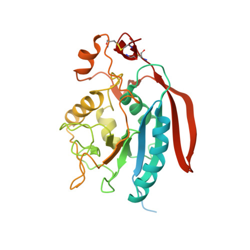
Crystal structure of the catalytic domain of Drosophila beta1,4-Galactosyltransferase-7.
Ramakrishnan, B., Qasba, P.K.(2010) J Biol Chem 285: 15619-15626
- PubMed: 20236943
- DOI: https://doi.org/10.1074/jbc.M109.099564
- Primary Citation of Related Structures:
3LW6 - PubMed Abstract:
The beta1,4-galactosyltransferase-7 (beta4Gal-T7) enzyme, one of seven members of the beta4Gal-T family, transfers in the presence of manganese Gal from UDP-Gal to an acceptor sugar (xylose) that is attached to a side chain hydroxyl group of Ser/Thr residues of proteoglycan proteins. It exhibits the least protein sequence similarity with the other family members, including the well studied family member beta4Gal-T1, which, in the presence of manganese, transfers Gal from UDP-Gal to GlcNAc. We report here the crystal structure of the catalytic domain of beta4Gal-T7 from Drosophila in the presence of manganese and UDP at 1.81 A resolution. In the crystal structure, a new manganese ion-binding motif (HXH) has been observed. Superposition of the crystal structures of beta4Gal-T7 and beta4Gal-T1 shows that the catalytic pocket and the substrate-binding sites in these proteins are similar. Compared with GlcNAc, xylose has a hydroxyl group (instead of an N-acetyl group) at C2 and lacks the CH(2)OH group at C5; thus, these protein structures show significant differences in their acceptor-binding site. Modeling of xylose in the acceptor-binding site of the beta4Gal-T7 crystal structure shows that the aromatic side chain of Tyr(177) interacts strongly with the C5 atom of xylose, causing steric hindrance to any additional group at C5. Because Drosophila Cd7 has a 73% protein sequence similarity to human Cd7, the present crystal structure offers a structure-based explanation for the mutations in human Cd7 that have been linked to Ehlers-Danlos syndrome.
Organizational Affiliation:
Structural Glycobiology Section, National Institutes of Health, Frederick, Maryland 21702; Basic Research Program, SAIC-Frederick, Inc., Center for Cancer Research Nanobiology Program, Center for Cancer Research, NCI, National Institutes of Health, Frederick, Maryland 21702.