


Crystal structure of ABC transporter, periplasmic substrate-binding protein SPO2066 from Silicibacter pomeroyi
Chang, C., Chhor, G., Clancy, S., Joachimiak, A.To be published.
Experimental Data Snapshot
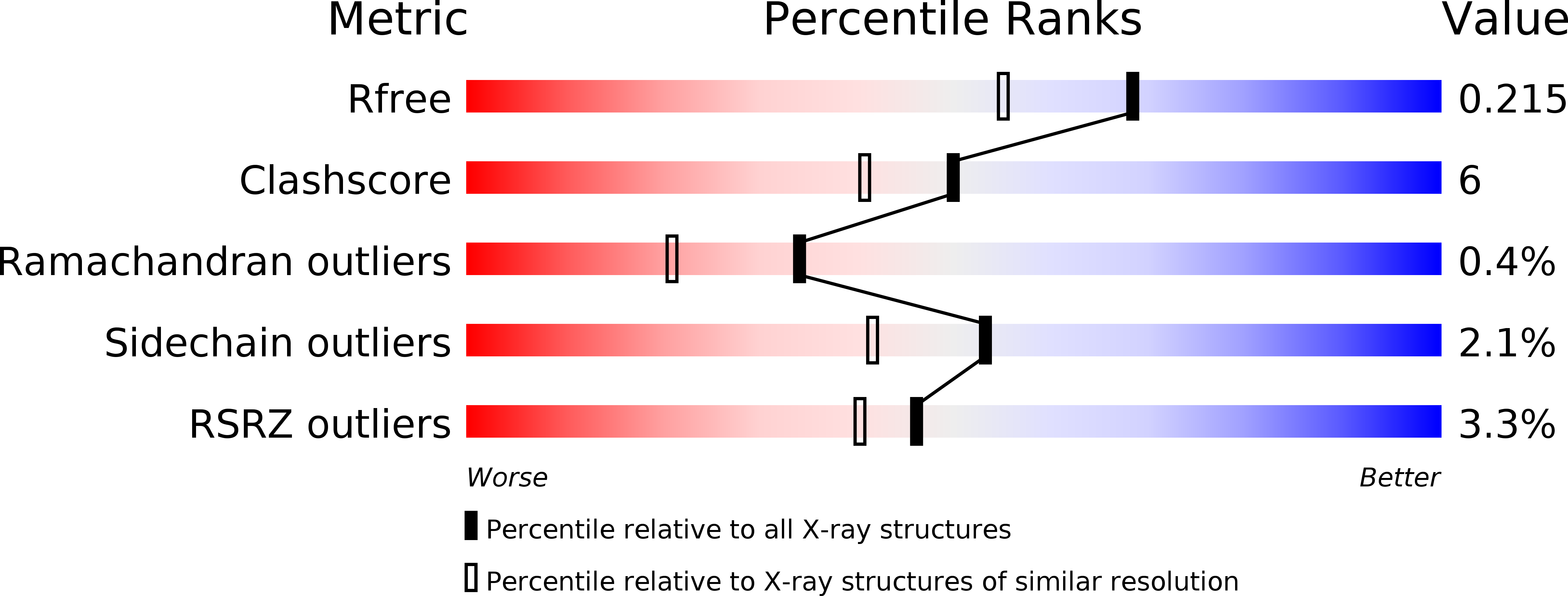
wwPDB Validation 3D Report Full Report
Entity ID: 1 | |||||
|---|---|---|---|---|---|
| Molecule | Chains | Sequence Length | Organism | Details | Image |
| ABC transporter, periplasmic substrate-binding protein | 258 | Ruegeria pomeroyi | Mutation(s): 0 Gene Names: SPO2066 | 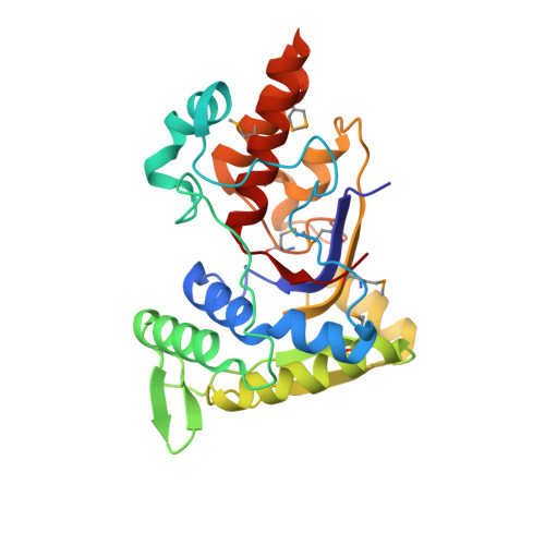 | |
UniProt | |||||
Find proteins for Q5LRQ9 (Ruegeria pomeroyi (strain ATCC 700808 / DSM 15171 / DSS-3)) Explore Q5LRQ9 Go to UniProtKB: Q5LRQ9 | |||||
Entity Groups | |||||
| Sequence Clusters | 30% Identity50% Identity70% Identity90% Identity95% Identity100% Identity | ||||
| UniProt Group | Q5LRQ9 | ||||
Sequence AnnotationsExpand | |||||
| |||||
| Ligands 4 Unique | |||||
|---|---|---|---|---|---|
| ID | Chains | Name / Formula / InChI Key | 2D Diagram | 3D Interactions | |
| PG5 Query on PG5 | P [auth B], Q [auth B] | 1-METHOXY-2-[2-(2-METHOXY-ETHOXY]-ETHANE C8 H18 O4 YFNKIDBQEZZDLK-UHFFFAOYSA-N |  | ||
| IOD Query on IOD | AA [auth D] BA [auth D] E [auth A] F [auth A] G [auth A] | IODIDE ION I XMBWDFGMSWQBCA-UHFFFAOYSA-M |  | ||
| GOL Query on GOL | H [auth A], O [auth B], W [auth C] | GLYCEROL C3 H8 O3 PEDCQBHIVMGVHV-UHFFFAOYSA-N |  | ||
| EDO Query on EDO | M [auth B], N [auth B], R [auth B], S [auth B], X [auth C] | 1,2-ETHANEDIOL C2 H6 O2 LYCAIKOWRPUZTN-UHFFFAOYSA-N |  | ||
| Modified Residues 1 Unique | |||||
|---|---|---|---|---|---|
| ID | Chains | Type | Formula | 2D Diagram | Parent |
| MSE Query on MSE | A, B, C, D | L-PEPTIDE LINKING | C5 H11 N O2 Se |  | MET |
| Length ( Å ) | Angle ( ˚ ) |
|---|---|
| a = 47.384 | α = 82.63 |
| b = 75.517 | β = 80.29 |
| c = 78.477 | γ = 77.42 |
| Software Name | Purpose |
|---|---|
| SBC-Collect | data collection |
| HKL-3000 | phasing |
| SHELXD | phasing |
| SHELXE | model building |
| MLPHARE | phasing |
| DM | model building |
| RESOLVE | model building |
| Coot | model building |
| ARP/wARP | model building |
| REFMAC | refinement |
| HKL-3000 | data reduction |
| HKL-3000 | data scaling |
| DM | phasing |
| RESOLVE | phasing |
RCSB PDB (citation) is hosted by
RCSB PDB is a member of the

