3LVB

Crystal structure of the Ferredoxin:NADP+ reductase from maize root at 1.7 angstroms - Test Set Withheld


Experimental Data Snapshot

  • Method: X-RAY DIFFRACTION
  • Resolution: 1.70 Å
  • R-Value Free: 0.223 
  • R-Value Work: 0.164 
  • R-Value Observed: 0.170 

wwPDB Validation   3D Report Full Report


Ligand Structure Quality Assessment 


This is version 1.2 of the entry. See complete history


Literature

Biochemical and crystallographic characterization of ferredoxin-NADP(+) reductase from nonphotosynthetic tissues

Aliverti, A.Faber, R.Finnerty, C.M.Ferioli, C.Pandini, V.Negri, A.Karplus, P.A.Zanetti, G.

(2001) Biochemistry 40: 14501-14508

  • DOI: https://doi.org/10.1021/bi011224c
  • Primary Citation of Related Structures:  
    1JB9, 3LVB

  • PubMed Abstract: 

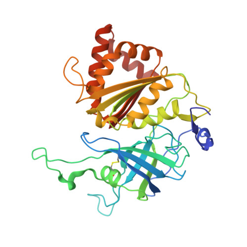
    Distinct forms of ferredoxin-NADP(+) reductase are expressed in photosynthetic and nonphotosynthetic plant tissues. Both enzymes catalyze electron transfer between NADP(H) and ferredoxin; whereas in leaves the enzyme transfers reducing equivalents from photoreduced ferredoxin to NADP(+) in photosynthesis, in roots it has the opposite physiological role, reducing ferredoxin at the expense of NADPH mainly for use in nitrate assimilation. Here, structural and kinetic properties of a nonphotosynthetic isoform were analyzed to define characteristics that may be related to tissue-specific function. Compared with spinach leaf ferredoxin-NADP(+) reductase, the recombinant corn root isoform showed a slightly altered absorption spectrum, a higher pI, a >30-fold higher affinity for NADP(+), greater susceptibility to limited proteolysis, and an approximately 20 mV more positive redox potential. The 1.7 A resolution crystal structure is very similar to the structures of ferredoxin-NADP(+) reductases from photosynthetic tissues. Four distinct structural features of this root ferredoxin-NADP(+) reductases are an alternate conformation of the bound FAD molecule, an alternate path for the amino-terminal extension, a disulfide bond in the FAD-binding domain, and changes in the surface that binds ferredoxin.


  • Organizational Affiliation

    Dipartimento di Fisiologia e Biochimica Generali, Università degli Studi di Milano, Via Celoria 26, 20133 Milano, Italy.


Macromolecules
Find similar proteins by:  (by identity cutoff)  |  3D Structure
Entity ID: 1
MoleculeChains Sequence LengthOrganismDetailsImage
Ferredoxin-NADP reductase311Zea maysMutation(s): 0 
EC: 1.18.1.2
UniProt
Find proteins for Q41736 (Zea mays)
Explore Q41736 
Go to UniProtKB:  Q41736
Entity Groups  
Sequence Clusters30% Identity50% Identity70% Identity90% Identity95% Identity100% Identity
UniProt GroupQ41736
Sequence Annotations
Expand
  • Reference Sequence
Small Molecules
Ligands 1 Unique
IDChains Name / Formula / InChI Key2D Diagram3D Interactions
FAD
Query on FAD

Download Ideal Coordinates CCD File 
B [auth A]FLAVIN-ADENINE DINUCLEOTIDE
C27 H33 N9 O15 P2
VWWQXMAJTJZDQX-UYBVJOGSSA-N
Experimental Data & Validation

Experimental Data

  • Method: X-RAY DIFFRACTION
  • Resolution: 1.70 Å
  • R-Value Free: 0.223 
  • R-Value Work: 0.164 
  • R-Value Observed: 0.170 
  • Space Group: P 32 2 1
Unit Cell:
Length ( Å )Angle ( ˚ )
a = 59.67α = 90
b = 59.67β = 90
c = 189.1γ = 120
Software Package:
Software NamePurpose
mrxmodel building
TNTrefinement
DENZOdata reduction
SCALEPACKdata scaling
mrxphasing

Structure Validation

View Full Validation Report


Ligand Structure Quality Assessment 


Entry History 

Deposition Data

Revision History  (Full details and data files)

  • Version 1.0: 2010-03-16
    Type: Initial release
  • Version 1.1: 2011-07-13
    Changes: Version format compliance
  • Version 1.2: 2023-09-06
    Changes: Data collection, Database references, Derived calculations, Refinement description