


Crystal structure of extragenic suppressor protein suhB from Bartonella henselae
Edwards, T.E., Abendroth, J., Sankaran, B., Seattle Structural Genomics Center for Infectious Disease (SSGCID)To be published.
Experimental Data Snapshot
wwPDB Validation 3D Report Full Report

Entity ID: 1 | |||||
|---|---|---|---|---|---|
| Molecule | Chains | Sequence Length | Organism | Details | Image |
| Extragenic suppressor protein suhB | 267 | Bartonella henselae | Mutation(s): 0 Gene Names: suhB, BH15030 | 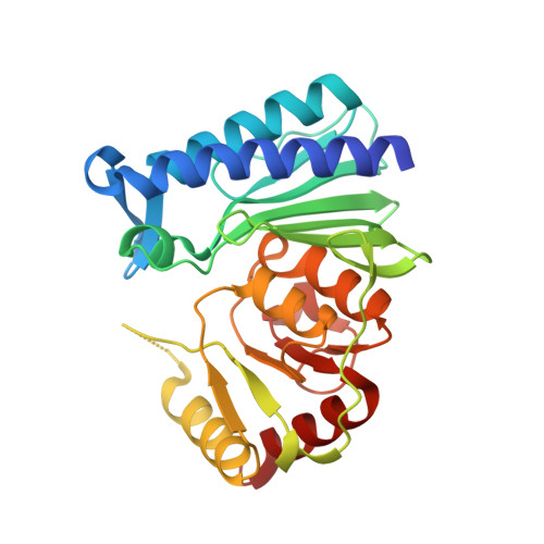 | |
UniProt | |||||
Find proteins for A0A0H3M6W8 (Bartonella henselae (strain ATCC 49882 / DSM 28221 / CCUG 30454 / Houston 1)) Explore A0A0H3M6W8 Go to UniProtKB: A0A0H3M6W8 | |||||
Entity Groups | |||||
| Sequence Clusters | 30% Identity50% Identity70% Identity90% Identity95% Identity100% Identity | ||||
| UniProt Group | A0A0H3M6W8 | ||||
Sequence AnnotationsExpand | |||||
| |||||
| Ligands 1 Unique | |||||
|---|---|---|---|---|---|
| ID | Chains | Name / Formula / InChI Key | 2D Diagram | 3D Interactions | |
| MG Query on MG | C [auth A], D [auth B], E [auth B] | MAGNESIUM ION Mg JLVVSXFLKOJNIY-UHFFFAOYSA-N |  | ||
| Length ( Å ) | Angle ( ˚ ) |
|---|---|
| a = 82.06 | α = 90 |
| b = 138.6 | β = 90 |
| c = 67.24 | γ = 90 |
| Software Name | Purpose |
|---|---|
| XSCALE | data scaling |
| PHASER | phasing |
| REFMAC | refinement |
| PDB_EXTRACT | data extraction |
| XDS | data reduction |
RCSB PDB (citation) is hosted by
RCSB PDB is a member of the

