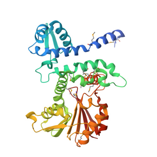
3LST

Crystal Structure of CalO1, Methyltransferase in Calicheamicin Biosynthesis, SAH bound form


Experimental Data Snapshot

  • Method: X-RAY DIFFRACTION
  • Resolution: 2.40 Å
  • R-Value Free: 0.251 
  • R-Value Work: 0.200 
  • R-Value Observed: 0.203 

wwPDB Validation   3D Report Full Report


Ligand Structure Quality Assessment 


This is version 1.2 of the entry. See complete history


Literature

Structural characterization of CalO1: a putative orsellinic acid methyltransferase in the calicheamicin-biosynthetic pathway.

Chang, A.Singh, S.Bingman, C.A.Thorson, J.S.Phillips, G.N.

(2011) Acta Crystallogr D Biol Crystallogr 67: 197-203

  • DOI: https://doi.org/10.1107/S090744491100360X
  • Primary Citation of Related Structures:  
    3LST

  • PubMed Abstract: 

    The X-ray structure determination at 2.4 Å resolution of the putative orsellinic acid C3 O-methyltransferase (CalO1) involved in calicheamicin biosynthesis is reported. Comparison of CalO1 with a homology model of the functionally related calicheamicin orsellinic acid C2 O-methyltransferase (CalO6) implicates several residues that are likely to contribute to the regiospecificity of alkylation. Consistent with the proposed requirement of an acyl-carrier-protein-bound substrate, this structural study also reveals structural determinants within CalO1 that are anticipated to accommodate an association with an acyl carrier protein.


  • Organizational Affiliation

    Department of Biochemistry, University of Wisconsin-Madison, Madison, Wisconsin 53706, USA.


Macromolecules
Find similar proteins by:  (by identity cutoff)  |  3D Structure
Entity ID: 1
MoleculeChains Sequence LengthOrganismDetailsImage
CalO1 Methyltransferase
A, B
348Micromonospora echinosporaMutation(s): 0 
Gene Names: calO1
UniProt
Find proteins for Q8KNE5 (Micromonospora echinospora)
Explore Q8KNE5 
Go to UniProtKB:  Q8KNE5
Entity Groups  
Sequence Clusters30% Identity50% Identity70% Identity90% Identity95% Identity100% Identity
UniProt GroupQ8KNE5
Sequence Annotations
Expand
  • Reference Sequence
Small Molecules
Experimental Data & Validation

Experimental Data

  • Method: X-RAY DIFFRACTION
  • Resolution: 2.40 Å
  • R-Value Free: 0.251 
  • R-Value Work: 0.200 
  • R-Value Observed: 0.203 
  • Space Group: C 2 2 21
Unit Cell:
Length ( Å )Angle ( ˚ )
a = 63.515α = 90
b = 93.603β = 90
c = 240.971γ = 90
Software Package:
Software NamePurpose
DENZOdata reduction
SCALEPACKdata scaling
SHARPphasing
PHENIXrefinement
PDB_EXTRACTdata extraction
HKL-2000data collection
HKL-2000data reduction
HKL-2000data scaling

Structure Validation

View Full Validation Report


Ligand Structure Quality Assessment 


Entry History 

Deposition Data

Revision History  (Full details and data files)

  • Version 1.0: 2010-03-02
    Type: Initial release
  • Version 1.1: 2011-07-13
    Changes: Version format compliance
  • Version 1.2: 2017-11-01
    Changes: Refinement description